2XAA | pdb_00002xaa

Alcohol dehydrogenase ADH-'A' from Rhodococcus ruber DSM 44541 at pH 8.5 in complex with NAD and butane-1,4-diol


Experimental Data Snapshot

  • Method: X-RAY DIFFRACTION
  • Resolution: 2.80 Å
  • R-Value Free: 
    0.258 (Depositor), 0.252 (DCC) 
  • R-Value Work: 
    0.188 (Depositor), 0.187 (DCC) 
  • R-Value Observed: 
    0.192 (Depositor) 

Starting Model: experimental
View more details

wwPDB Validation 3D Report Full Report

Validation slider image for 2XAA

Ligand Structure Quality Assessment 


This is version 1.3 of the entry. See complete history

Literature

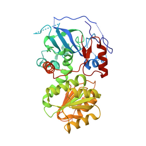
Structural Insights Into Substrate Specificity and Solvent Tolerance in Alcohol Dehydrogenase Adh-'A' from Rhodococcus Ruber Dsm 44541.

Karabec, M.Lyskowski, A.Tauber, K.C.Steinkellner, G.Kroutil, W.Grogan, G.Gruber, K.

(2010) Chem Commun (Camb) 46: 6314

  • DOI: https://doi.org/10.1039/c0cc00929f
  • Primary Citation Related Structures: 
    2XAA, 3JV7

  • PubMed Abstract: 

    The structure of the alcohol dehydrogenase ADH-'A' from Rhodococcus ruber reveals possible reasons for its remarkable tolerance to organic co-solvents and suggests new directions for structure-informed mutagenesis to produce enzymes of altered substrate specificity or improved selectivity.


  • Organizational Affiliation
    • Institute of Molecular Biosciences, University of Graz, Humboldtstrasse 50/3, A-8010 Graz, Austria.

Macromolecule Content 

  • Total Structure Weight: 144.33 kDa 
  • Atom Count: 10,078 
  • Modeled Residue Count: 1,364 
  • Deposited Residue Count: 1,380 
  • Unique protein chains: 1

Macromolecules

Find similar proteins by:|  3D Structure
Entity ID: 1
MoleculeChains  Sequence LengthOrganismDetailsImage
SECONDARY ALCOHOL DEHYDROGENASE
A, B, C, D
345Rhodococcus ruberMutation(s): 0 
EC: 1.1.1.1
UniProt
Find proteins for Q8KLT9 (Rhodococcus ruber)
Explore Q8KLT9 
Go to UniProtKB:  Q8KLT9
Entity Groups
Sequence Clusters30% Identity50% Identity70% Identity90% Identity95% Identity100% Identity
UniProt GroupQ8KLT9
Sequence Annotations
Expand
Reference Sequence

Small Molecules

Ligands 3 Unique
IDChains Name / Formula / InChI Key2D Diagram3D Interactions
NAD

Query on NAD


Download:Ideal Coordinates CCD File
E [auth A],
H [auth B],
L [auth C],
P [auth D]
NICOTINAMIDE-ADENINE-DINUCLEOTIDE
C21 H27 N7 O14 P2
BAWFJGJZGIEFAR-NNYOXOHSSA-N
BU1

Query on BU1


Download:Ideal Coordinates CCD File
J [auth B],
O [auth C],
R [auth D]
1,4-BUTANEDIOL
C4 H10 O2
WERYXYBDKMZEQL-UHFFFAOYSA-N
ZN

Query on ZN


Download:Ideal Coordinates CCD File
F [auth A]
G [auth A]
I [auth B]
K [auth B]
M [auth C]
F [auth A],
G [auth A],
I [auth B],
K [auth B],
M [auth C],
N [auth C],
Q [auth D],
S [auth D]
ZINC ION
Zn
PTFCDOFLOPIGGS-UHFFFAOYSA-N

Experimental Data & Validation

Experimental Data

  • Method: X-RAY DIFFRACTION
  • Resolution: 2.80 Å
  • R-Value Free:  0.258 (Depositor), 0.252 (DCC) 
  • R-Value Work:  0.188 (Depositor), 0.187 (DCC) 
  • R-Value Observed: 0.192 (Depositor) 
Space Group: P 1 21 1
Unit Cell:
Length ( Å )Angle ( ˚ )
a = 65.674α = 90
b = 105.1β = 91.27
c = 109.176γ = 90
Software Package:
Software NamePurpose
REFMACrefinement
HKL-2000data reduction
SCALEPACKdata scaling
MOLREPphasing

Structure Validation

View Full Validation Report


Ligand Structure Quality Assessment 


Entry History 

Deposition Data

Revision History  (Full details and data files)

  • Version 1.0: 2010-08-11
    Type: Initial release
  • Version 1.1: 2011-05-08
    Changes: Version format compliance
  • Version 1.2: 2011-07-13
    Changes: Version format compliance
  • Version 1.3: 2023-12-20
    Changes: Data collection, Database references, Derived calculations, Other, Refinement description