2X8H | pdb_00002x8h

Thioredoxin glutathione reductase from Schistosoma mansoni in complex with GSH


Experimental Data Snapshot

  • Method: X-RAY DIFFRACTION
  • Resolution: 1.90 Å
  • R-Value Free: 
    0.194 (Depositor), 0.195 (DCC) 
  • R-Value Work: 
    0.180 (Depositor), 0.181 (DCC) 
  • R-Value Observed: 
    0.181 (Depositor) 

Starting Model: experimental
View more details

wwPDB Validation 3D Report Full Report

Validation slider image for 2X8H

Ligand Structure Quality Assessment 


This is version 1.3 of the entry. See complete history

Literature

Mapping the Catalytic Cycle of Schistosoma Mansoni Thioredoxin Glutathione Reductase by X-Ray Crystallography

Angelucci, F.Dimastrogiovanni, D.Boumis, G.Brunori, M.Miele, A.E.Saccoccia, F.Bellelli, A.

(2010) J Biological Chem 285: 32557

  • DOI: https://doi.org/10.1074/jbc.M110.141960
  • Primary Citation Related Structures: 
    2X8C, 2X8G, 2X8H, 2X99

  • PubMed Abstract: 

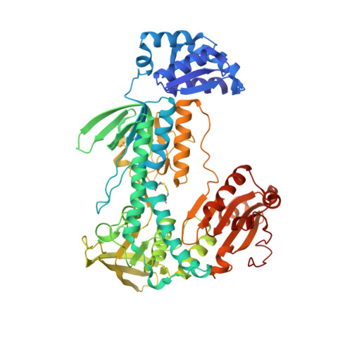
    Schistosomiasis is the second most widespread human parasitic disease. It is principally treated with one drug, praziquantel, that is administered to 100 million people each year; less sensitive strains of schistosomes are emerging. One of the most appealing drug targets against schistosomiasis is thioredoxin glutathione reductase (TGR). This natural chimeric enzyme is a peculiar fusion of a glutaredoxin domain with a thioredoxin selenocysteine (U)-containing reductase domain. Selenocysteine is located on a flexible C-terminal arm that is usually disordered in the available structures of the protein and is essential for the full catalytic activity of TGR. In this study, we dissect the catalytic cycle of Schistosoma mansoni TGR by structural and functional analysis of the U597C mutant. The crystallographic data presented herein include the following: the oxidized form (at 1.9 Å resolution); the NADPH- and GSH-bound forms (2.3 and 1.9 Å, respectively); and a different crystal form of the (partially) reduced enzyme (3.1 Å), showing the physiological dimer and the entire C terminus of one subunit. Whenever possible, we determined the rate constants for the interconversion between the different oxidation states of TGR by kinetic methods. By combining the crystallographic analysis with computer modeling, we were able to throw further light on the mechanism of action of S. mansoni TGR. In particular, we hereby propose the putative functionally relevant conformational change of the C terminus after the transfer of reducing equivalents from NADPH to the redox sites of the enzyme.


  • Organizational Affiliation
    • Department of Biochemical Sciences A. Rossi Fanelli, CNR Institute of Molecular Biology and Pathology and Istituto Pasteur-Fondazione Cenci Bolognetti, Sapienza University of Rome, P.le Aldo Moro 5, 00185 Rome, Italy.

Macromolecule Content 

  • Total Structure Weight: 67.19 kDa 
  • Atom Count: 5,009 
  • Modeled Residue Count: 588 
  • Deposited Residue Count: 598 
  • Unique protein chains: 1

Macromolecules

Find similar proteins by:|  3D Structure
Entity ID: 1
MoleculeChains  Sequence LengthOrganismDetailsImage
THIOREDOXIN GLUTATHIONE REDUCTASE598Schistosoma mansoniMutation(s): 0 
EC: 1.6.4.5 (PDB Primary Data), 1.8.1.9 (UniProt)
UniProt
Find proteins for Q962Y6 (Schistosoma mansoni)
Explore Q962Y6 
Go to UniProtKB:  Q962Y6
Entity Groups
Sequence Clusters30% Identity50% Identity70% Identity90% Identity95% Identity100% Identity
UniProt GroupQ962Y6
Sequence Annotations
Expand
Reference Sequence

Small Molecules

Ligands 7 Unique
IDChains Name / Formula / InChI Key2D Diagram3D Interactions
FAD

Query on FAD


Download:Ideal Coordinates CCD File
B [auth A]FLAVIN-ADENINE DINUCLEOTIDE
C27 H33 N9 O15 P2
VWWQXMAJTJZDQX-UYBVJOGSSA-N
GSH

Query on GSH


Download:Ideal Coordinates CCD File
C [auth A]Glutathione
C10 H17 N3 O6 S
RWSXRVCMGQZWBV-WDSKDSINSA-N
PG4

Query on PG4


Download:Ideal Coordinates CCD File
G [auth A]TETRAETHYLENE GLYCOL
C8 H18 O5
UWHCKJMYHZGTIT-UHFFFAOYSA-N
PGE

Query on PGE


Download:Ideal Coordinates CCD File
E [auth A],
F [auth A]
TRIETHYLENE GLYCOL
C6 H14 O4
ZIBGPFATKBEMQZ-UHFFFAOYSA-N
PEG

Query on PEG


Download:Ideal Coordinates CCD File
I [auth A],
J [auth A],
K [auth A],
L [auth A]
DI(HYDROXYETHYL)ETHER
C4 H10 O3
MTHSVFCYNBDYFN-UHFFFAOYSA-N
BME

Query on BME


Download:Ideal Coordinates CCD File
D [auth A]BETA-MERCAPTOETHANOL
C2 H6 O S
DGVVWUTYPXICAM-UHFFFAOYSA-N
CA

Query on CA


Download:Ideal Coordinates CCD File
H [auth A]CALCIUM ION
Ca
BHPQYMZQTOCNFJ-UHFFFAOYSA-N

Biologically Interesting Molecules (External Reference) 

1 Unique

Experimental Data & Validation

Experimental Data

  • Method: X-RAY DIFFRACTION
  • Resolution: 1.90 Å
  • R-Value Free:  0.194 (Depositor), 0.195 (DCC) 
  • R-Value Work:  0.180 (Depositor), 0.181 (DCC) 
  • R-Value Observed: 0.181 (Depositor) 
Space Group: C 1 2 1
Unit Cell:
Length ( Å )Angle ( ˚ )
a = 141.576α = 90
b = 102.885β = 112.62
c = 59.112γ = 90
Software Package:
Software NamePurpose
REFMACrefinement
MOSFLMdata reduction
SCALAdata scaling
PDBSETphasing

Structure Validation

View Full Validation Report


Ligand Structure Quality Assessment 


Entry History 

Deposition Data

Revision History  (Full details and data files)

  • Version 1.0: 2010-07-21
    Type: Initial release
  • Version 1.1: 2011-12-07
    Changes: Atomic model, Derived calculations, Non-polymer description, Version format compliance
  • Version 1.2: 2019-01-30
    Changes: Data collection, Experimental preparation
  • Version 1.3: 2023-12-20
    Changes: Data collection, Database references, Derived calculations, Other, Refinement description