2X5I | pdb_00002x5i

Crystal structure echovirus 7


Experimental Data Snapshot

  • Method: X-RAY DIFFRACTION
  • Resolution: 3.10 Å
  • R-Value Work: 
    0.288 (Depositor), 0.282 (DCC) 
  • R-Value Observed: 
    0.288 (Depositor) 

Starting Model: experimental
View more details

wwPDB Validation 3D Report Full Report

Validation slider image for 2X5I

Ligand Structure Quality Assessment 


This is version 1.4 of the entry. See complete history

Literature

Interaction of Decay-Accelerating Factor with Echovirus 7.

Plevka, P.Hafenstein, S.Harris, K.G.Cifuente, J.O.Zhang, Y.Bowman, V.D.Chipman, P.R.Bator, C.M.Lin, F.Medof, M.E.Rossmann, M.G.

(2010) J Virol 84: 12665

  • DOI: https://doi.org/10.1128/JVI.00837-10
  • Primary Citation Related Structures: 
    2X5I, 3IYP

  • PubMed Abstract: 

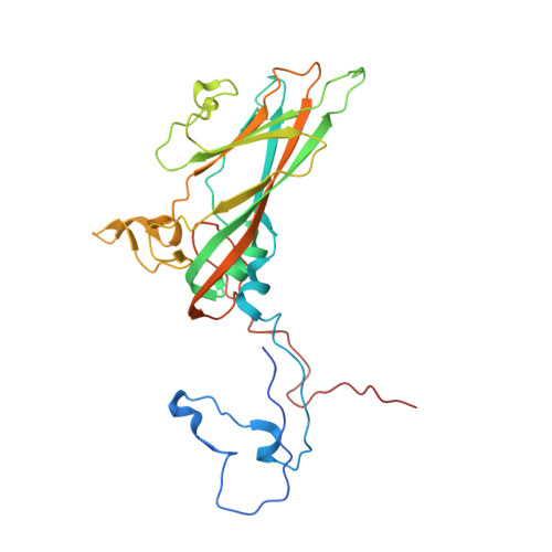
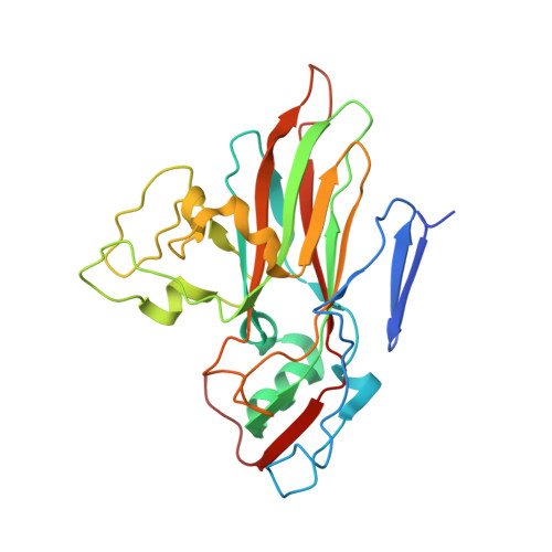
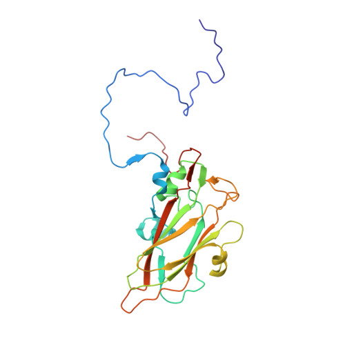
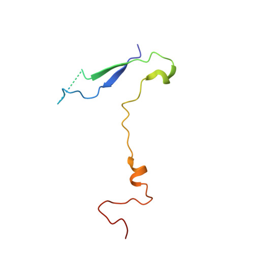
    Echovirus 7 (EV7) belongs to the Enterovirus genus within the family Picornaviridae. Many picornaviruses use IgG-like receptors that bind in the viral canyon and are required to initiate viral uncoating during infection. However, in addition, some of the enteroviruses use an alternative or additional receptor that binds outside the canyon. Decay-accelerating factor (DAF) has been identified as a cellular receptor for EV7. The crystal structure of EV7 has been determined to 3.1-Å resolution and used to interpret the 7.2-Å-resolution cryo-electron microscopy reconstruction of EV7 complexed with DAF. Each DAF binding site on EV7 is near a 2-fold icosahedral symmetry axis, which differs from the binding site of DAF on the surface of coxsackievirus B3, indicating that there are independent evolutionary processes by which DAF was selected as a picornavirus accessory receptor. This suggests that there is an advantage for these viruses to recognize DAF during the initial process of infection.


  • Organizational Affiliation
    • Department of Biological Sciences, Purdue University, 915 W. State Street, West Lafayette, IN 47907-2054, USA.

Macromolecule Content 

  • Total Structure Weight: 96.41 kDa 
  • Atom Count: 6,540 
  • Modeled Residue Count: 828 
  • Deposited Residue Count: 860 
  • Unique protein chains: 4

Macromolecules

Find similar proteins by:|  3D Structure
Entity ID: 1
MoleculeChains  Sequence LengthOrganismDetailsImage
VP1292Echovirus E7Mutation(s): 0 
UniProt
Find proteins for Q6W9E5 (Echovirus E7)
Explore Q6W9E5 
Go to UniProtKB:  Q6W9E5
Entity Groups
Sequence Clusters30% Identity50% Identity70% Identity90% Identity95% Identity100% Identity
UniProt GroupQ6W9E5
Sequence Annotations
Expand
Reference Sequence
Find similar proteins by:|  3D Structure
Entity ID: 2
MoleculeChains  Sequence LengthOrganismDetailsImage
VP2260Echovirus E7Mutation(s): 0 
UniProt
Find proteins for Q6W9E5 (Echovirus E7)
Explore Q6W9E5 
Go to UniProtKB:  Q6W9E5
Entity Groups
Sequence Clusters30% Identity50% Identity70% Identity90% Identity95% Identity100% Identity
UniProt GroupQ6W9E5
Sequence Annotations
Expand
Reference Sequence
Find similar proteins by:|  3D Structure
Entity ID: 3
MoleculeChains  Sequence LengthOrganismDetailsImage
VP3238Echovirus E7Mutation(s): 0 
UniProt
Find proteins for Q6W9E5 (Echovirus E7)
Explore Q6W9E5 
Go to UniProtKB:  Q6W9E5
Entity Groups
Sequence Clusters30% Identity50% Identity70% Identity90% Identity95% Identity100% Identity
UniProt GroupQ6W9E5
Sequence Annotations
Expand
Reference Sequence
Find similar proteins by:|  3D Structure
Entity ID: 4
MoleculeChains  Sequence LengthOrganismDetailsImage
VP470Echovirus E7Mutation(s): 0 
UniProt
Find proteins for Q6W9E5 (Echovirus E7)
Explore Q6W9E5 
Go to UniProtKB:  Q6W9E5
Entity Groups
Sequence Clusters30% Identity50% Identity70% Identity90% Identity95% Identity100% Identity
UniProt GroupQ6W9E5
Sequence Annotations
Expand
Reference Sequence

Small Molecules

Ligands 1 Unique
IDChains Name / Formula / InChI Key2D Diagram3D Interactions
DAO

Query on DAO


Download:Ideal Coordinates CCD File
E [auth A]LAURIC ACID
C12 H24 O2
POULHZVOKOAJMA-UHFFFAOYSA-N

Experimental Data & Validation

Experimental Data

  • Method: X-RAY DIFFRACTION
  • Resolution: 3.10 Å
  • R-Value Work:  0.288 (Depositor), 0.282 (DCC) 
  • R-Value Observed: 0.288 (Depositor) 
Space Group: P 1
Unit Cell:
Length ( Å )Angle ( ˚ )
a = 297.1α = 119
b = 297.7β = 100.1
c = 300.6γ = 108.4
Software Package:
Software NamePurpose
HKL-2000data reduction
HKL-2000data scaling
GLRFphasing
CNSphasing
CNSrefinement

Structure Validation

View Full Validation Report


Ligand Structure Quality Assessment 


Entry History 

Deposition Data

Revision History  (Full details and data files)

  • Version 1.0: 2010-12-22
    Type: Initial release
  • Version 1.1: 2011-05-08
    Changes: Version format compliance
  • Version 1.2: 2011-07-13
    Changes: Version format compliance
  • Version 1.3: 2019-05-08
    Changes: Data collection, Experimental preparation, Other
  • Version 1.4: 2023-12-20
    Changes: Data collection, Database references, Derived calculations, Other, Refinement description