2X25 | pdb_00002x25

Free acetyl-CypA orthorhombic form


Experimental Data Snapshot

  • Method: X-RAY DIFFRACTION
  • Resolution: 1.20 Å
  • R-Value Free: 
    0.156 (Depositor), 0.168 (DCC) 
  • R-Value Work: 
    0.126 (Depositor), 0.143 (DCC) 
  • R-Value Observed: 
    0.128 (Depositor) 

wwPDB Validation 3D Report Full Report

Validation slider image for 2X25

This is version 1.3 of the entry. See complete history

Literature

Acetylation Regulates Cyclophilin a Catalysis, Immunosuppression and HIV Isomerization.

Lammers, M.Neumann, H.Chin, J.W.James, L.C.

(2010) Nat Chem Biol 6: 331

  • DOI: https://doi.org/10.1038/nchembio.342
  • Primary Citation Related Structures: 
    2X25, 2X2A, 2X2C, 2X2D

  • PubMed Abstract: 

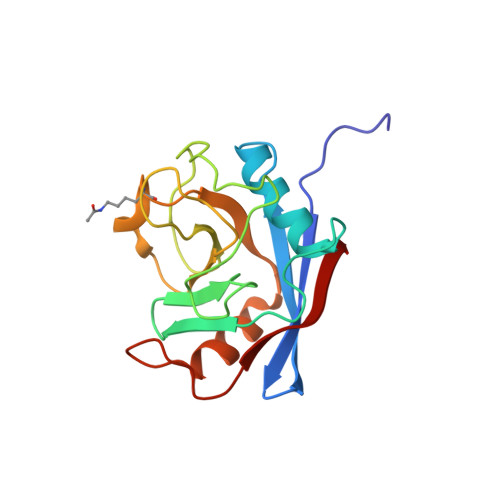
    Cyclophilin A (CypA) is a ubiquitous cis-trans prolyl isomerase with key roles in immunity and viral infection. CypA suppresses T-cell activation through cyclosporine complexation and is required for effective HIV-1 replication in host cells. We show that CypA is acetylated in diverse human cell lines and use a synthetically evolved acetyllysyl-tRNA synthetase/tRNA(CUA) pair to produce recombinant acetylated CypA in Escherichia coli. We determined atomic-resolution structures of acetylated CypA and its complexes with cyclosporine and HIV-1 capsid. Acetylation markedly inhibited CypA catalysis of cis to trans isomerization and stabilized cis rather than trans forms of the HIV-1 capsid. Furthermore, CypA acetylation antagonized the immunosuppressive effects of cyclosporine by inhibiting the sequential steps of cyclosporine binding and calcineurin inhibition. Our results reveal that acetylation regulates key functions of CypA in immunity and viral infection and provide a general set of mechanisms by which acetylation modulates interactions to regulate cell function.


  • Organizational Affiliation
    • Medical Research Council Laboratory of Molecular Biology, Protein and Nucleic Acid Chemistry Division, Cambridge, UK.

Macromolecule Content 

  • Total Structure Weight: 18.64 kDa 
  • Atom Count: 1,744 
  • Modeled Residue Count: 168 
  • Deposited Residue Count: 169 
  • Unique protein chains: 1

Macromolecules

Find similar proteins by:|  3D Structure
Entity ID: 1
MoleculeChains  Sequence LengthOrganismDetailsImage
PEPTIDYL-PROLYL CIS-TRANS ISOMERASE AA [auth B]169Homo sapiensMutation(s): 0 
EC: 5.2.1.8
UniProt & NIH Common Fund Data Resources
Find proteins for P62937 (Homo sapiens)
Explore P62937 
Go to UniProtKB:  P62937
PHAROS:  P62937
GTEx:  ENSG00000196262 
Entity Groups
Sequence Clusters30% Identity50% Identity70% Identity90% Identity95% Identity100% Identity
UniProt GroupP62937
Sequence Annotations
Expand
Reference Sequence

Small Molecules

Modified Residues  1 Unique
IDChains TypeFormula2D DiagramParent
ALY
Query on ALY
A [auth B]L-PEPTIDE LINKINGC8 H16 N2 O3LYS

Experimental Data & Validation

Experimental Data

  • Method: X-RAY DIFFRACTION
  • Resolution: 1.20 Å
  • R-Value Free:  0.156 (Depositor), 0.168 (DCC) 
  • R-Value Work:  0.126 (Depositor), 0.143 (DCC) 
  • R-Value Observed: 0.128 (Depositor) 
Space Group: P 21 21 21
Unit Cell:
Length ( Å )Angle ( ˚ )
a = 42.222α = 90
b = 52.404β = 90
c = 89.236γ = 90
Software Package:
Software NamePurpose
REFMACrefinement

Structure Validation

View Full Validation Report


Entry History 

Deposition Data

Revision History  (Full details and data files)

  • Version 1.0: 2010-03-23
    Type: Initial release
  • Version 1.1: 2011-06-09
    Changes: Version format compliance
  • Version 1.2: 2011-07-13
    Changes: Version format compliance
  • Version 1.3: 2024-11-13
    Changes: Data collection, Database references, Derived calculations, Other, Structure summary