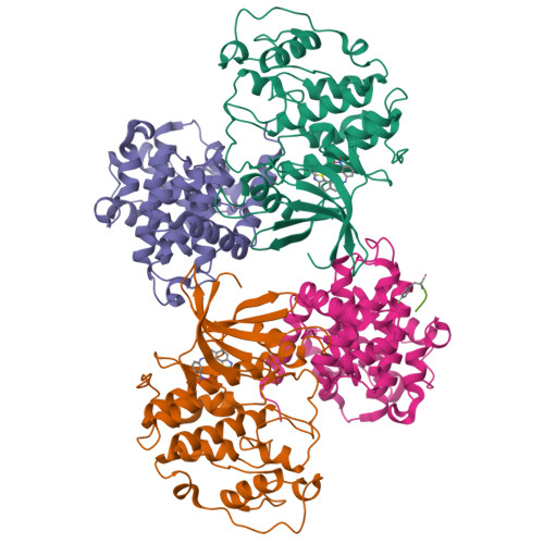
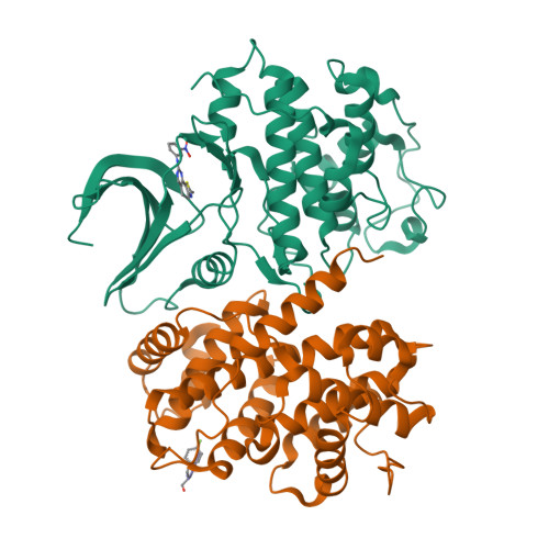
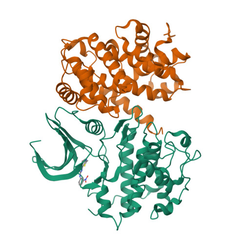
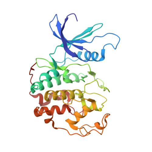
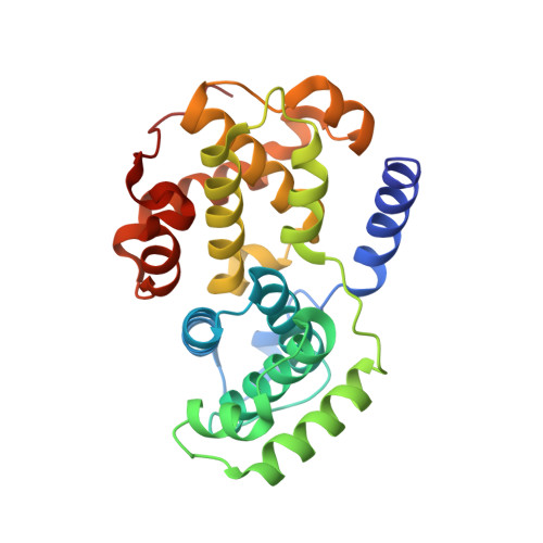
2X1N

Truncation and Optimisation of Peptide Inhibitors of CDK2, Cyclin A Through Structure Guided Design


Experimental Data Snapshot

  • Method: X-RAY DIFFRACTION
  • Resolution: 2.75 Å
  • R-Value Free: 0.255 
  • R-Value Work: 0.200 
  • R-Value Observed: 0.201 

Starting Model: experimental
View more details

wwPDB Validation   3D Report Full Report


Ligand Structure Quality Assessment 


This is version 1.3 of the entry. See complete history


Literature

Design, Synthesis, and Evaluation of 2-Methyl- and 2-Amino-N-Aryl-4,5-Dihydrothiazolo[4,5-H]Quinazolin-8-Amines as Ring-Constrained 2-Anilino-4-(Thiazol-5-Yl)Pyrimidine Cyclin-Dependent Kinase Inhibitors.

Mcintyre, N.A.Mcinnes, C.Griffiths, G.Barnett, A.L.Kontopidis, G.Slawin, A.M.Z.Jackson, W.Thomas, M.Zheleva, D.I.Wang, S.Blake, D.G.Westwood, N.J.Fischer, P.M.

(2010) J Med Chem 53: 2136

  • DOI: https://doi.org/10.1021/jm901660c
  • Primary Citation of Related Structures:  
    2X1N

  • PubMed Abstract: 

    Following the recent discovery and development of 2-anilino-4-(thiazol-5-yl)pyrimidine cyclin dependent kinase (CDK) inhibitors, a program was initiated to evaluate related ring-constrained analogues, specifically, 2-methyl- and 2-amino-N-aryl-4,5-dihydrothiazolo[4,5-h]quinazolin-8-amines for inhibition of CDKs. Here we report the rational design, synthesis, structure-activity relationships (SARs), and cellular mode-of-action profile of these second generation CDK inhibitors. Many of the analogues from this chemical series inhibit CDKs with very low nanomolar K(i) values. The most potent compound reported in this study inhibits CDK2 with an IC(50) of 0.7 nM ([ATP] = 100 microM). Furthermore, an X-ray crystal structure of 2-methyl-N-(3-(nitro)phenyl)-4,5-dihydrothiazolo[4,5-h]quinazolin-8-amine (11g), a representative from the chemical series in complex with cyclin A-CDK2, is reported, confirming the design rationale and expected binding mode within the CDK2 ATP binding pocket.


  • Organizational Affiliation

    School of Chemistry and Biomedical Sciences Research Complex, University of St. Andrews, North Haugh, St Andrews, Fife KY16 9ST, UK.


Macromolecules
Find similar proteins by:  (by identity cutoff)  |  3D Structure
Entity ID: 1
MoleculeChains Sequence LengthOrganismDetailsImage
CELL DIVISION PROTEIN KINASE 2
A, C
298Homo sapiensMutation(s): 0 
EC: 2.7.1.37 (PDB Primary Data), 2.7.11.22 (UniProt)
UniProt & NIH Common Fund Data Resources
Find proteins for P24941 (Homo sapiens)
Explore P24941 
Go to UniProtKB:  P24941
PHAROS:  P24941
GTEx:  ENSG00000123374 
Entity Groups  
Sequence Clusters30% Identity50% Identity70% Identity90% Identity95% Identity100% Identity
UniProt GroupP24941
Sequence Annotations
Expand
  • Reference Sequence
Find similar proteins by:  (by identity cutoff)  |  3D Structure
Entity ID: 2
MoleculeChains Sequence LengthOrganismDetailsImage
CYCLIN-A2
B, D
261Homo sapiensMutation(s): 0 
UniProt & NIH Common Fund Data Resources
Find proteins for P20248 (Homo sapiens)
Explore P20248 
Go to UniProtKB:  P20248
PHAROS:  P20248
GTEx:  ENSG00000145386 
Entity Groups  
Sequence Clusters30% Identity50% Identity70% Identity90% Identity95% Identity100% Identity
UniProt GroupP20248
Sequence Annotations
Expand
  • Reference Sequence

Find similar proteins by:  Sequence   |   3D Structure  

Entity ID: 3
MoleculeChains Sequence LengthOrganismDetailsImage
ACE-LEU-ASN-PFF-NH2E [auth H]5synthetic constructMutation(s): 0 
Sequence Annotations
Expand
  • Reference Sequence
Small Molecules
Ligands 1 Unique
IDChains Name / Formula / InChI Key2D Diagram3D Interactions
X1N
Query on X1N

Download Ideal Coordinates CCD File 
F [auth A],
G [auth C]
2-METHYL-N-[(1Z)-3-NITROCYCLOHEXA-2,4-DIEN-1-YLIDENE]-4,5-DIHYDRO[1,3]THIAZOLO[4,5-H]QUINAZOLIN-8-AMINE
C16 H13 N5 O2 S
CMWWYSDXIZEJPA-ODLFYWEKSA-N
Modified Residues  1 Unique
IDChains TypeFormula2D DiagramParent
PFF
Query on PFF
E [auth H]L-PEPTIDE LINKINGC9 H10 F N O2PHE
Experimental Data & Validation

Experimental Data

  • Method: X-RAY DIFFRACTION
  • Resolution: 2.75 Å
  • R-Value Free: 0.255 
  • R-Value Work: 0.200 
  • R-Value Observed: 0.201 
  • Space Group: P 21 21 21
Unit Cell:
Length ( Å )Angle ( ˚ )
a = 74.558α = 90
b = 114.257β = 90
c = 157.277γ = 90
Software Package:
Software NamePurpose
REFMACrefinement
MOSFLMdata reduction
SCALAdata scaling
MOLREPphasing

Structure Validation

View Full Validation Report


Ligand Structure Quality Assessment 


Entry History 

Revision History  (Full details and data files)

  • Version 1.0: 2010-02-16
    Type: Initial release
  • Version 1.1: 2016-12-14
    Changes: Source and taxonomy, Version format compliance
  • Version 1.2: 2017-03-29
    Changes: Other
  • Version 1.3: 2023-12-20
    Changes: Data collection, Database references, Derived calculations, Other, Refinement description