2X17 | pdb_00002x17

The X-ray structure of Ferritin from Pyrococcus furiosus loaded with Ag(I)


Experimental Data Snapshot

  • Method: X-RAY DIFFRACTION
  • Resolution: 3.10 Å
  • R-Value Free: 
    0.283 (Depositor), 0.269 (DCC) 
  • R-Value Work: 
    0.260 (Depositor), 0.248 (DCC) 
  • R-Value Observed: 
    0.261 (Depositor) 

Starting Model: experimental
View more details

wwPDB Validation 3D Report Full Report

Validation slider image for 2X17

This is version 1.2 of the entry. See complete history

Literature

Silver Ion Incorporation and Nanoparticle Formation Inside the Cavity of Pyrococcus Furiosus Ferritin: Structural and Size-Distribution Analyses.

Kasyutich, O.Ilari, A.Fiorillo, A.Tatchev, D.Hoell, A.Ceci, P.

(2010) J Am Chem Soc 132: 3621

  • DOI: https://doi.org/10.1021/ja910918b
  • Primary Citation Related Structures: 
    2X17

  • PubMed Abstract: 

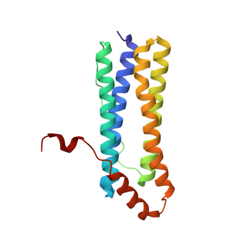
    Highly symmetrical protein cage architectures from three different iron storage proteins, heavy and light human ferritin chains (HuHFt and HuLFt) and ferritin from the hyperthemophilic bacterium Pyrococcus furiosus (PfFt), have been used as models for understanding the molecular basis of silver ion deposition and metal core formation inside the protein cavity. Biomineralization using protein cavities is an important issue for the fabrication of biometamaterials under mild synthetic conditions. Silver nanoparticles (AgNPs) were produced with high yields within PfFt but not within HuHFt and HuLFt. To explain the molecular basis of silver incorporation, the X-ray crystal structure of Ag-containing PfFt has been solved. This is the first structure of a silver containing ferritin reported to date, and it revealed the presence of specific binding and nucleation sites of Ag(I) that are not conserved in other ferritin templates. The AgNP encapsulated by PfFt were further characterized by the combined use of different physical-chemical techniques. These showed that the AgNPs are endowed with a narrow size distribution (2.1 +/- 0.4 nm), high stability in water solution at millimolar concentration, and high thermal stability. These properties make the AgNP obtained within PftFt exploitable for a range of applications, in fields as diverse as catalysis in water, preparation of metamaterials, and in vivo diagnosis and antibacterial or tumor therapy.


  • Organizational Affiliation
    • University of Bristol, Physics Department, HH Wills Physics Laboratory, Tyndall Avenues, Bristol, BS8 1TL, UK.

Macromolecule Content 

  • Total Structure Weight: 488.46 kDa 
  • Atom Count: 34,042 
  • Modeled Residue Count: 4,110 
  • Deposited Residue Count: 4,152 
  • Unique protein chains: 1

Macromolecules

Find similar proteins by:|  3D Structure
Entity ID: 1
MoleculeChains  Sequence LengthOrganismDetailsImage
PUTATIVE FERRITIN HOMOLOG173Pyrococcus furiosusMutation(s): 0 
UniProt
Find proteins for Q8U2T8 (Pyrococcus furiosus (strain ATCC 43587 / DSM 3638 / JCM 8422 / Vc1))
Explore Q8U2T8 
Go to UniProtKB:  Q8U2T8
Entity Groups
Sequence Clusters30% Identity50% Identity70% Identity90% Identity95% Identity100% Identity
UniProt GroupQ8U2T8
Sequence Annotations
Expand
Reference Sequence

Small Molecules

Ligands 1 Unique
IDChains Name / Formula / InChI Key2D Diagram3D Interactions
AG

Query on AG


Download:Ideal Coordinates CCD File
AA [auth 1]
AB [auth P]
BA [auth 1]
BB [auth Q]
CA [auth 2]
AA [auth 1],
AB [auth P],
BA [auth 1],
BB [auth Q],
CA [auth 2],
CB [auth R],
DA [auth 3],
DB [auth Y],
EA [auth 3],
EB [auth Z],
FA [auth 3],
GA [auth 4],
HA [auth 4],
IA [auth 5],
JA [auth 5],
KA [auth 6],
LA [auth 6],
MA [auth 7],
NA [auth 8],
OA [auth 9],
PA [auth G],
QA [auth G],
RA [auth H],
SA [auth I],
TA [auth J],
UA [auth K],
VA [auth L],
WA [auth M],
XA [auth M],
Y [auth 0],
YA [auth N],
Z [auth 1],
ZA [auth O]
SILVER ION
Ag
FOIXSVOLVBLSDH-UHFFFAOYSA-N

Experimental Data & Validation

Experimental Data

  • Method: X-RAY DIFFRACTION
  • Resolution: 3.10 Å
  • R-Value Free:  0.283 (Depositor), 0.269 (DCC) 
  • R-Value Work:  0.260 (Depositor), 0.248 (DCC) 
  • R-Value Observed: 0.261 (Depositor) 
Space Group: P 41
Unit Cell:
Length ( Å )Angle ( ˚ )
a = 157.681α = 90
b = 157.681β = 90
c = 246.437γ = 90
Software Package:
Software NamePurpose
REFMACrefinement
DENZOdata reduction
SCALEPACKdata scaling
MOLREPphasing

Structure Validation

View Full Validation Report


Entry History 

Deposition Data

Revision History  (Full details and data files)

  • Version 1.0: 2010-04-21
    Type: Initial release
  • Version 1.1: 2011-07-27
    Changes: Refinement description, Version format compliance
  • Version 1.2: 2023-12-20
    Changes: Data collection, Database references, Derived calculations, Other, Refinement description