2WW8 | pdb_00002ww8

Structure of the pilus adhesin (RrgA) from Streptococcus pneumoniae


Experimental Data Snapshot

  • Method: X-RAY DIFFRACTION
  • Resolution: 1.90 Å
  • R-Value Free: 
    0.235 (Depositor), 0.217 (DCC) 
  • R-Value Work: 
    0.195 (Depositor), 0.216 (DCC) 
  • R-Value Observed: 
    0.197 (Depositor) 

wwPDB Validation 3D Report Full Report

Validation slider image for 2WW8

This is version 1.5 of the entry. See complete history

Literature

Structural Basis of Host Cell Recognition by the Pilus Adhesin from Streptococcus Pneumoniae

Izore, T.Contreras-Martel, C.El-Mortaji, L.Manzano, C.Terrasse, R.Vernet, T.Di-Guilmi, A.M.Dessen, A.

(2010) Structure 18: 106

  • DOI: https://doi.org/10.1016/j.str.2009.10.019
  • Primary Citation Related Structures: 
    2WW8

  • PubMed Abstract: 

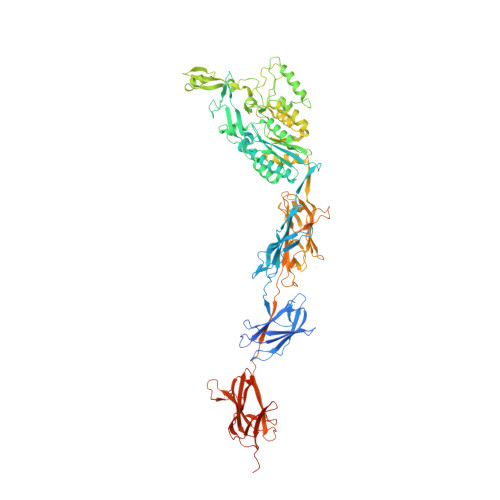
    Pili are fibrous virulence factors associated directly to the bacterial surface that play critical roles in adhesion and recognition of host cell receptors. The human pathogen Streptococcus pneumoniae carries a single pilus-related adhesin (RrgA) that is key for infection establishment and provides protection from bacterial challenge in animal infection models, but details of these roles remain unclear. Here we report the high-resolution crystal structure of RrgA, a 893-residue elongated macromolecule whose fold contains four domains presenting both eukaryotic and prokaryotic origins. RrgA harbors an integrin I collagen-recognition domain decorated with two inserted "arms" that fold into a positively charged cradle, as well as three "stalk-forming" domains. We show by site-specific mutagenesis, mass spectrometry, and thermal shift assays that intradomain isopeptide bonds play key roles in stabilizing RrgA's stalk. The high sequence similarity between RrgA and its homologs in other Gram-positive microorganisms suggests common strategies for ECM recognition and immune evasion.


  • Organizational Affiliation
    • Institut de Biologie Structurale Jean-Pierre Ebel, UMR 5075 (CEA, CNRS, UJF), Grenoble, France.

Macromolecule Content 

  • Total Structure Weight: 100.43 kDa 
  • Atom Count: 7,294 
  • Modeled Residue Count: 815 
  • Deposited Residue Count: 893 
  • Unique protein chains: 1

Macromolecules

Find similar proteins by:|  3D Structure
Entity ID: 1
MoleculeChains  Sequence LengthOrganismDetailsImage
CELL WALL SURFACE ANCHOR FAMILY PROTEIN893Streptococcus pneumoniaeMutation(s): 0 
UniProt
Find proteins for A0A0H2UNT6 (Streptococcus pneumoniae serotype 4 (strain ATCC BAA-334 / TIGR4))
Explore A0A0H2UNT6 
Go to UniProtKB:  A0A0H2UNT6
Entity Groups
Sequence Clusters30% Identity50% Identity70% Identity90% Identity95% Identity100% Identity
UniProt GroupA0A0H2UNT6
Sequence Annotations
Expand
Reference Sequence

Experimental Data & Validation

Experimental Data

  • Method: X-RAY DIFFRACTION
  • Resolution: 1.90 Å
  • R-Value Free:  0.235 (Depositor), 0.217 (DCC) 
  • R-Value Work:  0.195 (Depositor), 0.216 (DCC) 
  • R-Value Observed: 0.197 (Depositor) 
Space Group: P 21 21 21
Unit Cell:
Length ( Å )Angle ( ˚ )
a = 51.081α = 90
b = 82.7β = 90
c = 299.338γ = 90
Software Package:
Software NamePurpose
REFMACrefinement
XDSdata reduction
XSCALEdata scaling
autoSHARPphasing

Structure Validation

View Full Validation Report


Entry History 

Deposition Data

Revision History  (Full details and data files)

  • Version 1.0: 2010-01-19
    Type: Initial release
  • Version 1.1: 2011-07-13
    Changes: Advisory, Version format compliance
  • Version 1.2: 2013-03-06
    Changes: Data collection
  • Version 1.3: 2024-05-08
    Changes: Data collection, Database references, Derived calculations, Other
  • Version 1.4: 2024-06-05
    Changes: Advisory, Derived calculations
  • Version 1.5: 2024-10-16
    Changes: Structure summary