2WV0 | pdb_00002wv0

Crystal structure of the GntR-HutC family member YvoA from Bacillus subtilis


Experimental Data Snapshot

  • Method: X-RAY DIFFRACTION
  • Resolution: 2.40 Å
  • R-Value Free: 
    0.255 (Depositor), 0.261 (DCC) 
  • R-Value Work: 
    0.199 (Depositor), 0.258 (DCC) 
  • R-Value Observed: 
    0.205 (Depositor) 

wwPDB Validation 3D Report Full Report

Validation slider image for 2WV0

This is version 1.3 of the entry. See complete history

Literature

Insight Into the Induction Mechanism of the Gntr/Hutc Bacterial Transcription Regulator Yvoa

Resch, M.Schiltz, E.Titgemeyer, F.Muller, Y.A.

(2010) Nucleic Acids Res 38: 2485

  • DOI: https://doi.org/10.1093/nar/gkp1191
  • Primary Citation Related Structures: 
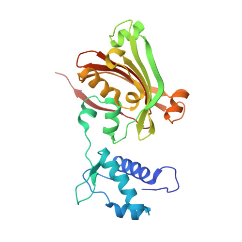
    2WV0

  • PubMed Abstract: 

    YvoA is a GntR/HutC transcription regulator from Bacillus subtilis implicated in the regulation of genes from the N-acetylglucosamine-degrading pathway. Its 2.4-A crystal structure reveals a homodimeric assembly with each monomer displaying a two-domain fold. The C-terminal domain, which binds the effector N-acetylglucosamine-6-phosphate, adopts a chorismate lyase fold, whereas the N-terminal domain contains a winged helix-turn-helix DNA-binding domain. Isothermal titration calorimetry and site-directed mutagenesis revealed that the effector-binding site in YvoA coincides with the active site of related chorismate lyase from Escherichia coli. The characterization of the DNA- and effector-binding properties of two disulfide-bridged mutants that lock YvoA in two distinct conformational states provides for the first time detailed insight into the allosteric mechanism through which effector binding modulates DNA binding and, thereby regulates transcription in a representative GntR/HutC family member. Central to this allosteric coupling mechanism is a loop-to-helix transition with the dipole of the newly formed helix pointing toward the phosphate of the effector. This transition goes in hand with the emergence of internal symmetry in the effector-binding domain and, in addition, leads to a 122 degrees rotation of the DNA-binding domains that is best described as a jumping-jack-like motion.


  • Organizational Affiliation
    • Lehrstuhl für Biotechnik, Department of Biology, Friedrich-Alexander University Erlangen-Nuremberg, D-91052 Erlangen, Germany.

Macromolecule Content 

  • Total Structure Weight: 276.85 kDa 
  • Atom Count: 18,689 
  • Modeled Residue Count: 2,378 
  • Deposited Residue Count: 2,430 
  • Unique protein chains: 1

Macromolecules

Find similar proteins by:|  3D Structure
Entity ID: 1
MoleculeChains  Sequence LengthOrganismDetailsImage
HTH-TYPE TRANSCRIPTIONAL REPRESSOR YVOA
A, B, C, D, E
A, B, C, D, E, F, G, H, I, J
243Bacillus subtilisMutation(s): 0 
UniProt
Find proteins for O34817 (Bacillus subtilis (strain 168))
Explore O34817 
Go to UniProtKB:  O34817
Entity Groups
Sequence Clusters30% Identity50% Identity70% Identity90% Identity95% Identity100% Identity
UniProt GroupO34817
Sequence Annotations
Expand
Reference Sequence

Small Molecules

Ligands 1 Unique
IDChains Name / Formula / InChI Key2D Diagram3D Interactions
SO4

Query on SO4


Download:Ideal Coordinates CCD File
K [auth A]
L [auth A]
M [auth B]
N [auth B]
O [auth C]
K [auth A],
L [auth A],
M [auth B],
N [auth B],
O [auth C],
P [auth C],
Q [auth D],
R [auth D],
S [auth E],
T [auth F],
U [auth F],
V [auth G],
W [auth G],
X [auth I],
Y [auth I]
SULFATE ION
O4 S
QAOWNCQODCNURD-UHFFFAOYSA-L

Experimental Data & Validation

Experimental Data

  • Method: X-RAY DIFFRACTION
  • Resolution: 2.40 Å
  • R-Value Free:  0.255 (Depositor), 0.261 (DCC) 
  • R-Value Work:  0.199 (Depositor), 0.258 (DCC) 
  • R-Value Observed: 0.205 (Depositor) 
Space Group: C 1 2 1
Unit Cell:
Length ( Å )Angle ( ˚ )
a = 208.41α = 90
b = 137.08β = 94.97
c = 120.19γ = 90
Software Package:
Software NamePurpose
REFMACrefinement
XDSdata reduction
XSCALEdata scaling
autoSHARPphasing

Structure Validation

View Full Validation Report


Entry History 

Deposition Data

Revision History  (Full details and data files)

  • Version 1.0: 2010-01-12
    Type: Initial release
  • Version 1.1: 2011-05-08
    Changes: Version format compliance
  • Version 1.2: 2011-07-13
    Changes: Version format compliance
  • Version 1.3: 2024-05-08
    Changes: Data collection, Database references, Derived calculations, Other