2WU8 | pdb_00002wu8

Structural studies of phosphoglucose isomerase from Mycobacterium tuberculosis H37Rv


Experimental Data Snapshot

  • Method: X-RAY DIFFRACTION
  • Resolution: 2.25 Å
  • R-Value Free: 
    0.223 (Depositor), 0.216 (DCC) 
  • R-Value Work: 
    0.182 (Depositor), 0.173 (DCC) 
  • R-Value Observed: 
    0.183 (Depositor) 

Starting Model: experimental
View more details

wwPDB Validation 3D Report Full Report

Validation slider image for 2WU8

This is version 1.3 of the entry. See complete history

Literature

Structural Studies of Phosphoglucose Isomerase from Mycobacterium Tuberculosis H37Rv

Anand, K.Mathur, D.Anant, A.Garg, L.C.

(2010) Acta Crystallogr Sect F Struct Biol Cryst Commun 66: 490

  • DOI: https://doi.org/10.1107/S1744309110011656
  • Primary Citation Related Structures: 
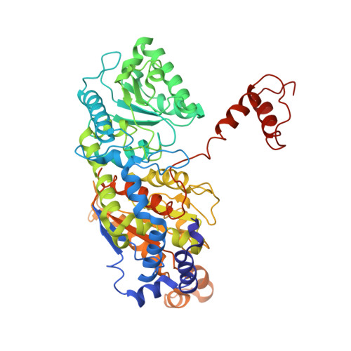
    2WU8

  • PubMed Abstract: 

    Phosphoglucose isomerase (PGI) plays a key role in both glycolysis and gluconeogenesis inside the cell, whereas outside the cell it exhibits cytokine properties. PGI is also known to act as an autocrine motility factor, a neuroleukin agent and a differentiation and maturation mediator. Here, the first crystal structure of PGI from Mycobacterium tuberculosis H37Rv (Mtb) is reported. The structure was refined at 2.25 A resolution and revealed the presence of one molecule in the asymmetric unit with two globular domains. As known previously, the active site of Mtb PGI contains conserved residues including Glu356, Glu216 and His387 (where His387 is from the neighbouring molecule). The crystal structure of Mtb PGI was observed to be rather more similar to human PGI than other nonbacterial PGIs, with only a few differences being detected in the loops, arm and hook regions of the human and Mtb PGIs, suggesting that the M. tuberculosis enzyme uses the same enzyme mechanism.


  • Organizational Affiliation
    • European Molecular Biology Laboratory Heidelberg, Structural and Computational Biology Unit, Meyerhof Strasse 1, D-69117 Heidelberg, Germany. anand@embl.de,lalit@nii.res.in

Macromolecule Content 

  • Total Structure Weight: 59.78 kDa 
  • Atom Count: 4,483 
  • Modeled Residue Count: 549 
  • Deposited Residue Count: 549 
  • Unique protein chains: 1

Macromolecules

Find similar proteins by:|  3D Structure
Entity ID: 1
MoleculeChains  Sequence LengthOrganismDetailsImage
GLUCOSE-6-PHOSPHATE ISOMERASE549Mycobacterium tuberculosis H37RvMutation(s): 0 
EC: 5.3.1.9
UniProt
Find proteins for P9WN69 (Mycobacterium tuberculosis (strain ATCC 25618 / H37Rv))
Explore P9WN69 
Go to UniProtKB:  P9WN69
Entity Groups
Sequence Clusters30% Identity50% Identity70% Identity90% Identity95% Identity100% Identity
UniProt GroupP9WN69
Sequence Annotations
Expand
Reference Sequence

Experimental Data & Validation

Experimental Data

  • Method: X-RAY DIFFRACTION
  • Resolution: 2.25 Å
  • R-Value Free:  0.223 (Depositor), 0.216 (DCC) 
  • R-Value Work:  0.182 (Depositor), 0.173 (DCC) 
  • R-Value Observed: 0.183 (Depositor) 
Space Group: I 21 21 21
Unit Cell:
Length ( Å )Angle ( ˚ )
a = 109.7α = 90
b = 118.5β = 90
c = 137.6γ = 90
Software Package:
Software NamePurpose
PHENIXrefinement
XDSdata reduction
XSCALEdata scaling
PHASERphasing

Structure Validation

View Full Validation Report


Entry History 

Deposition Data

  • Released Date: 2009-10-27 
  • Deposition Author(s): Anand, K.

Revision History  (Full details and data files)

  • Version 1.0: 2009-10-27
    Type: Initial release
  • Version 1.1: 2011-05-08
    Changes: Version format compliance
  • Version 1.2: 2011-07-13
    Changes: Version format compliance
  • Version 1.3: 2023-12-20
    Changes: Data collection, Database references, Derived calculations, Other, Refinement description