2WPW | pdb_00002wpw

Tandem GNAT protein from the clavulanic acid biosynthesis pathway (without AcCoA)


Experimental Data Snapshot

  • Method: X-RAY DIFFRACTION
  • Resolution: 2.38 Å
  • R-Value Free: 
    0.266 (Depositor), 0.263 (DCC) 
  • R-Value Work: 
    0.209 (Depositor), 0.214 (DCC) 
  • R-Value Observed: 
    0.212 (Depositor) 

wwPDB Validation 3D Report Full Report

Validation slider image for 2WPW

Ligand Structure Quality Assessment 


This is version 1.3 of the entry. See complete history

Literature

Crystallographic and mass spectrometric analyses of a tandem GNAT protein from the clavulanic acid biosynthesis pathway.

Iqbal, A.Arunlanantham, H.Brown, T.Chowdhury, R.Clifton, I.J.Kershaw, N.J.Hewitson, K.S.McDonough, M.A.Schofield, C.J.

(2010) Proteins 78: 1398-1407

  • DOI: https://doi.org/10.1002/prot.22653
  • Primary Citation Related Structures: 
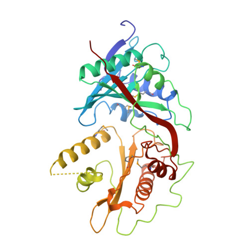
    2WPW, 2WPX

  • PubMed Abstract: 

    (3R,5R)-Clavulanic acid (CA) is a clinically important inhibitor of Class A beta-lactamases. Sequence comparisons suggest that orf14 of the clavulanic acid biosynthesis gene cluster encodes for an acetyl transferase (CBG). Crystallographic studies reveal CBG to be a member of the emerging structural subfamily of tandem Gcn5-related acetyl transferase (GNAT) proteins. Two crystal forms (C2 and P2(1) space groups) of CBG were obtained; in both forms one molecule of acetyl-CoA (AcCoA) was bound to the N-terminal GNAT domain, with the C-terminal domain being unoccupied by a ligand. Mass spectrometric analyzes on CBG demonstrate that, in addition to one strongly bound AcCoA molecule, a second acyl-CoA molecule can bind to CBG. Succinyl-CoA and myristoyl-CoA displayed the strongest binding to the "second" CoA binding site, which is likely in the C-terminal GNAT domain. Analysis of the CBG structures, together with those of other tandem GNAT proteins, suggest that the AcCoA in the N-terminal GNAT domain plays a structural role whereas the C-terminal domain is more likely to be directly involved in acetyl transfer. The available crystallographic and mass spectrometric evidence suggests that binding of the second acyl-CoA occurs preferentially to monomeric rather than dimeric CBG. The N-terminal AcCoA binding site and the proposed C-terminal acyl-CoA binding site of CBG are compared with acyl-CoA binding sites of other tandem and single domain GNAT proteins.


  • Organizational Affiliation
    • University of Oxford, United Kingdom.

Macromolecule Content 

  • Total Structure Weight: 149.9 kDa 
  • Atom Count: 11,259 
  • Modeled Residue Count: 1,304 
  • Deposited Residue Count: 1,356 
  • Unique protein chains: 1

Macromolecules

Find similar proteins by:|  3D Structure
Entity ID: 1
MoleculeChains  Sequence LengthOrganismDetailsImage
ORF14
A, B, C, D
339Streptomyces clavuligerusMutation(s): 0 
EC: 2.3.1
UniProt
Find proteins for Q8KRB5 (Streptomyces clavuligerus)
Explore Q8KRB5 
Go to UniProtKB:  Q8KRB5
Entity Groups
Sequence Clusters30% Identity50% Identity70% Identity90% Identity95% Identity100% Identity
UniProt GroupQ8KRB5
Sequence Annotations
Expand
Reference Sequence

Experimental Data & Validation

Experimental Data

  • Method: X-RAY DIFFRACTION
  • Resolution: 2.38 Å
  • R-Value Free:  0.266 (Depositor), 0.263 (DCC) 
  • R-Value Work:  0.209 (Depositor), 0.214 (DCC) 
  • R-Value Observed: 0.212 (Depositor) 
Space Group: P 1 21 1
Unit Cell:
Length ( Å )Angle ( ˚ )
a = 75.449α = 90
b = 93.501β = 97.86
c = 126.43γ = 90
Software Package:
Software NamePurpose
REFMACrefinement
DENZOdata reduction
SCALEPACKdata scaling
SOLVEphasing

Structure Validation

View Full Validation Report


Ligand Structure Quality Assessment 


Entry History 

Deposition Data

Revision History  (Full details and data files)

  • Version 1.0: 2009-12-29
    Type: Initial release
  • Version 1.1: 2011-11-09
    Changes: Database references, Derived calculations, Non-polymer description, Other, Refinement description, Version format compliance
  • Version 1.2: 2018-03-28
    Changes: Database references
  • Version 1.3: 2024-11-13
    Changes: Data collection, Database references, Derived calculations, Other, Structure summary