2WL7 | pdb_00002wl7

Crystal structure of the PSD93 PDZ1 domain


Experimental Data Snapshot

  • Method: X-RAY DIFFRACTION
  • Resolution: 2.03 Å
  • R-Value Free: 
    0.208 (Depositor), 0.166 (DCC) 
  • R-Value Work: 
    0.164 (Depositor), 0.164 (DCC) 
  • R-Value Observed: 
    0.169 (Depositor) 

Starting Model: experimental
View more details

wwPDB Validation 3D Report Full Report

Validation slider image for 2WL7

This is version 1.3 of the entry. See complete history

Literature

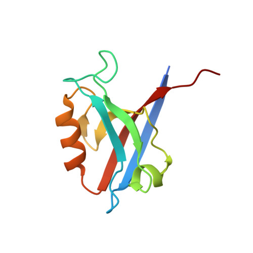
Structure of the First Pdz Domain of Human Psd-93.

Fiorentini, M.Nielsen, A.K.Kristensen, O.Kastrup, J.S.Gajhede, M.

(2009) Acta Crystallogr Sect F Struct Biol Cryst Commun 65: 1254

  • DOI: https://doi.org/10.1107/S1744309109043267
  • Primary Citation Related Structures: 
    2WL7

  • PubMed Abstract: 

    The crystal structure of the PDZ1 domain of human PSD-93 has been determined to 2.0 A resolution. The PDZ1 domain forms a crystallographic trimer that is also predicted to be stable in solution. The main contributions to the stabilization of the trimer seem to arise from interactions involving the PDZ1-PDZ2 linker region at the extreme C-terminus of PDZ1, implying that the oligomerization that is observed is not of biological significance in full-length PSD-93. Comparison of the structures of the binding cleft of PSD-93 PDZ1 with the previously reported structures of PSD-93 PDZ2 and PDZ3 as well as of the closely related human PSD-95 PDZ1 shows that they are very similar in terms of amino-acid composition. However, the cleft is significantly narrower in PSD-95. This could be part of the basis of peptide selectivity between PSD-93 PDZ1 and PSD-95 PDZ1.


  • Organizational Affiliation
    • Biostructural Research, Department of Medicinal Chemistry, Faculty of Pharmaceutical Sciences, University of Copenhagen, Universitetsparken 2, DK-2100 Copenhagen, Denmark.

Macromolecule Content 

  • Total Structure Weight: 11.28 kDa 
  • Atom Count: 882 
  • Modeled Residue Count: 96 
  • Deposited Residue Count: 102 
  • Unique protein chains: 1

Macromolecules

Find similar proteins by:|  3D Structure
Entity ID: 1
MoleculeChains  Sequence LengthOrganismDetailsImage
DISKS LARGE HOMOLOG 2102Mus musculusMutation(s): 0 
EC: 2.7.4.8
UniProt & NIH Common Fund Data Resources
Find proteins for Q91XM9 (Mus musculus)
Explore Q91XM9 
Go to UniProtKB:  Q91XM9
IMPC:  MGI:1344351
Entity Groups
Sequence Clusters30% Identity50% Identity70% Identity90% Identity95% Identity100% Identity
UniProt GroupQ91XM9
Sequence Annotations
Expand
Reference Sequence

Experimental Data & Validation

Experimental Data

  • Method: X-RAY DIFFRACTION
  • Resolution: 2.03 Å
  • R-Value Free:  0.208 (Depositor), 0.166 (DCC) 
  • R-Value Work:  0.164 (Depositor), 0.164 (DCC) 
  • R-Value Observed: 0.169 (Depositor) 
Space Group: H 3
Unit Cell:
Length ( Å )Angle ( ˚ )
a = 85.198α = 90
b = 85.198β = 90
c = 45.515γ = 120
Software Package:
Software NamePurpose
PHENIXrefinement
MOSFLMdata reduction
SCALAdata scaling
PHASERphasing

Structure Validation

View Full Validation Report


Entry History 

Deposition Data

Revision History  (Full details and data files)

  • Version 1.0: 2010-01-19
    Type: Initial release
  • Version 1.1: 2011-05-08
    Changes: Version format compliance
  • Version 1.2: 2011-07-13
    Changes: Version format compliance
  • Version 1.3: 2023-12-13
    Changes: Data collection, Database references, Derived calculations, Other, Refinement description