2WKX | pdb_00002wkx

Crystal structure of the native E. coli zinc amidase AmiD


Experimental Data Snapshot

  • Method: X-RAY DIFFRACTION
  • Resolution: 1.80 Å
  • R-Value Free: 
    0.184 (Depositor), 0.182 (DCC) 
  • R-Value Work: 
    0.161 (Depositor), 0.159 (DCC) 
  • R-Value Observed: 
    0.163 (Depositor) 

Starting Model: experimental
View more details

wwPDB Validation 3D Report Full Report

Validation slider image for 2WKX

This is version 1.2 of the entry. See complete history

Literature

Specific Structural Features of the N-Acetylmuramoyl-L-Alanine Amidase Amid from Escherichia Coli and Mechanistic Implications for Enzymes of This Family.

Kerff, F.Petrella, S.Mercier, F.Sauvage, E.Herman, R.Pennartz, A.Zervosen, A.Luxen, A.Frere, J.M.Joris, B.Charlier, P.

(2010) J Mol Biology 397: 249

  • DOI: https://doi.org/10.1016/j.jmb.2009.12.038
  • Primary Citation Related Structures: 
    2BH7, 2WKX, 3D2Y, 3D2Z

  • PubMed Abstract: 

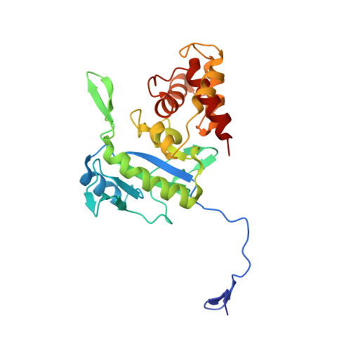
    AmiD is the fifth identified N-acetylmuramoyl-L-alanine zinc amidase of Escherichia coli. This periplasmic lipoprotein is anchored in the outer membrane and has a broad specificity. AmiD is capable of cleaving the intact peptidoglycan (PG) as well as soluble fragments containing N-acetylmuramic acid regardless of the presence of an anhydro form or not, unlike the four other amidases, AmiA, AmiB, AmiC, and AmpD, which have some specificity. AmiD function is, however, not clearly established but it could be part of the enzymatic machinery involved in the PG turnover in E. coli. We solved three structures of the E. coli zinc amidase AmiD devoid of its lipidic anchorage: the holoenzyme, the apoenzyme in complex with the substrate anhydro-N-acetylmuramic-acid-L-Ala-gamma-d-Glu-L-Lys, and the holoenzyme in complex with the L-Ala-gamma-D-Glu-L-Lys peptide, the product of the hydrolysis of this substrate by AmiD. The AmiD structure shows a relatively flexible N-terminal extension that allows an easy reach of the PG by the enzyme inserted into the outer membrane. The C-terminal domain provides a potential extended geometrical complementarity to the substrate. AmiD shares a common fold with AmpD, the bacteriophage T7 lysozyme, and the PG recognition proteins, which are receptor proteins involved in the innate immune responses of a wide range of organisms. Analysis of the different structures reveals the similarity between the catalytic mechanism of zinc amidases of the AmiD family and the thermolysin-related zinc peptidases.


  • Organizational Affiliation
    • Centre d'Ingénierie des Protéines, University of Liège, Allée du 6 Août 17, Bât B5a, 4000 Liège, Belgium.

Macromolecule Content 

  • Total Structure Weight: 29.88 kDa 
  • Atom Count: 2,351 
  • Modeled Residue Count: 257 
  • Deposited Residue Count: 261 
  • Unique protein chains: 1

Macromolecules

Find similar proteins by:|  3D Structure
Entity ID: 1
MoleculeChains  Sequence LengthOrganismDetailsImage
N-ACETYLMURAMOYL-L-ALANINE AMIDASE AMID261Escherichia coli K-12Mutation(s): 0 
EC: 3.5.1.28
UniProt
Find proteins for P75820 (Escherichia coli (strain K12))
Explore P75820 
Go to UniProtKB:  P75820
Entity Groups
Sequence Clusters30% Identity50% Identity70% Identity90% Identity95% Identity100% Identity
UniProt GroupP75820
Sequence Annotations
Expand
Reference Sequence

Small Molecules

Ligands 3 Unique
IDChains Name / Formula / InChI Key2D Diagram3D Interactions
GOL

Query on GOL


Download:Ideal Coordinates CCD File
E [auth A],
F [auth A],
G [auth A],
H [auth A]
GLYCEROL
C3 H8 O3
PEDCQBHIVMGVHV-UHFFFAOYSA-N
ZN

Query on ZN


Download:Ideal Coordinates CCD File
B [auth A]ZINC ION
Zn
PTFCDOFLOPIGGS-UHFFFAOYSA-N
CL

Query on CL


Download:Ideal Coordinates CCD File
C [auth A],
D [auth A]
CHLORIDE ION
Cl
VEXZGXHMUGYJMC-UHFFFAOYSA-M

Experimental Data & Validation

Experimental Data

  • Method: X-RAY DIFFRACTION
  • Resolution: 1.80 Å
  • R-Value Free:  0.184 (Depositor), 0.182 (DCC) 
  • R-Value Work:  0.161 (Depositor), 0.159 (DCC) 
  • R-Value Observed: 0.163 (Depositor) 
Space Group: P 61 2 2
Unit Cell:
Length ( Å )Angle ( ˚ )
a = 88.99α = 90
b = 88.99β = 90
c = 183.923γ = 120
Software Package:
Software NamePurpose
REFMACrefinement
MOSFLMdata reduction
SCALAdata scaling
AMoREphasing

Structure Validation

View Full Validation Report


Entry History 

Deposition Data

Revision History  (Full details and data files)

  • Version 1.0: 2010-01-12
    Type: Initial release
  • Version 1.1: 2011-07-13
    Changes: Advisory, Version format compliance
  • Version 1.2: 2023-12-13
    Changes: Data collection, Database references, Derived calculations, Other, Refinement description