2WJW | pdb_00002wjw

Crystal structure of the human ionotropic glutamate receptor GluR2 ATD region at 1.8 A resolution


Experimental Data Snapshot

  • Method: X-RAY DIFFRACTION
  • Resolution: 1.80 Å
  • R-Value Free: 
    0.217 (Depositor), 0.216 (DCC) 
  • R-Value Work: 
    0.181 (Depositor), 0.183 (DCC) 
  • R-Value Observed: 
    0.183 (Depositor) 

wwPDB Validation   3D Report Full Report


Ligand Structure Quality Assessment 

Created with Raphaël 2.3.0Worse 01 BetterLigand structure goodness of fit to experimental dataBest fitted NAGClick on this verticalbar to view details

This is version 1.4 of the entry. See complete history


Literature

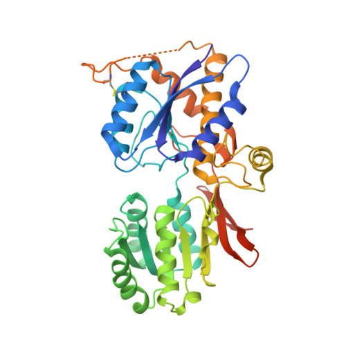
Crystal Structure of the Glur2 Amino-Terminal Domain Provides Insights Into the Architecture and Assembly of Ionotropic Glutamate Receptors.

Clayton, A.Siebold, C.Gilbert, R.J.C.Sutton, G.C.Harlos, K.Mcilhinney, R.A.J.Jones, E.Y.Aricescu, A.R.

(2009) J Mol Biology 392: 1125

  • DOI: https://doi.org/10.1016/j.jmb.2009.07.082
  • Primary Citation of Related Structures:  
    2WJW, 2WJX

  • PubMed Abstract: 

    Ionotropic glutamate receptors are functionally diverse but have a common architecture, including the 400-residue amino-terminal domain (ATD). We report a 1.8-A resolution crystal structure of human GluR2-ATD. This dimeric structure provides a mechanism for how the ATDs can drive receptor assembly and subtype-restricted composition. Lattice contacts in a 4.1-A resolution crystal form reveal a tetrameric (dimer-dimer) arrangement consistent with previous cellular and cryo-electron microscopic data for full-length AMPA receptors.


  • Organizational Affiliation

    Division of Structural Biology, Wellcome Trust Centre for Human Genetics, University of Oxford, Roosevelt Drive, Oxford, UK.


Macromolecules
Find similar proteins by:  (by identity cutoff)  |  3D Structure
Entity ID: 1
MoleculeChains Sequence LengthOrganismDetailsImage
GLUTAMATE RECEPTOR 2388Homo sapiensMutation(s): 0 
UniProt & NIH Common Fund Data Resources
Find proteins for P42262 (Homo sapiens)
Explore P42262 
Go to UniProtKB:  P42262
PHAROS:  P42262
GTEx:  ENSG00000120251 
Entity Groups  
Sequence Clusters30% Identity50% Identity70% Identity90% Identity95% Identity100% Identity
UniProt GroupP42262
Glycosylation
Glycosylation Sites: 1Go to GlyGen: P42262-1
Sequence Annotations
Expand
  • Reference Sequence
Experimental Data & Validation

Experimental Data

  • Method: X-RAY DIFFRACTION
  • Resolution: 1.80 Å
  • R-Value Free:  0.217 (Depositor), 0.216 (DCC) 
  • R-Value Work:  0.181 (Depositor), 0.183 (DCC) 
  • R-Value Observed: 0.183 (Depositor) 
Space Group: I 2 2 2
Unit Cell:
Length ( Å )Angle ( ˚ )
a = 50.977α = 90
b = 122.455β = 90
c = 138.852γ = 90
Software Package:
Software NamePurpose
PHENIXrefinement
HKL-2000data reduction
SCALEPACKdata scaling
autoSHARPphasing

Structure Validation

View Full Validation Report


Ligand Structure Quality Assessment 

Created with Raphaël 2.3.0Worse 01 BetterLigand structure goodness of fit to experimental dataBest fitted NAGClick on this verticalbar to view details

Entry History 

Deposition Data

Revision History  (Full details and data files)

  • Version 1.0: 2009-08-18
    Type: Initial release
  • Version 1.1: 2011-05-08
    Changes: Version format compliance
  • Version 1.2: 2011-07-13
    Changes: Version format compliance
  • Version 1.3: 2020-07-29
    Type: Remediation
    Reason: Carbohydrate remediation
    Changes: Data collection, Derived calculations, Other, Structure summary
  • Version 1.4: 2024-10-16
    Changes: Data collection, Database references, Structure summary