2WIY | pdb_00002wiy

Cytochrome P450 XplA heme domain P21212


Experimental Data Snapshot

  • Method: X-RAY DIFFRACTION
  • Resolution: 1.49 Å
  • R-Value Free: 
    0.178 (Depositor), 0.178 (DCC) 
  • R-Value Work: 
    0.147 (Depositor), 0.148 (DCC) 
  • R-Value Observed: 
    0.149 (Depositor) 

Starting Model: experimental
View more details

wwPDB Validation 3D Report Full Report

Validation slider image for 2WIY

Ligand Structure Quality Assessment 


This is version 1.3 of the entry. See complete history

Literature

The 1.5-A Structure of Xpla-Heme, an Unusual Cytochrome P450 Heme Domain that Catalyzes Reductive Biotransformation of Royal Demolition Explosive.

Sabbadin, F.Jackson, R.Haider, K.Tampi, G.Turkenburg, J.P.Hart, S.Bruce, N.C.Grogan, G.

(2009) J Biological Chem 284: 28467

  • DOI: https://doi.org/10.1074/jbc.M109.031559
  • Primary Citation Related Structures: 
    2WIV, 2WIY

  • PubMed Abstract: 

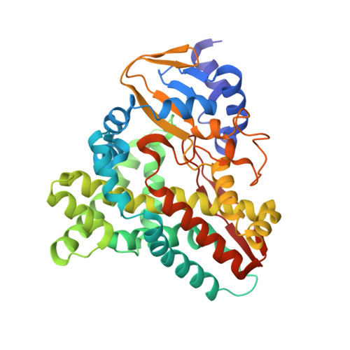
    XplA is a cytochrome P450 of unique structural organization, consisting of a heme-domain that is C-terminally fused to its native flavodoxin redox partner. XplA, along with flavodoxin reductase XplB, has been shown to catalyze the breakdown of the nitramine explosive and pollutant hexahydro-1,3,5-trinitro-1,3,5-triazine (royal demolition explosive) by reductive denitration. The structure of the heme domain of XplA (XplA-heme) has been solved in two crystal forms: as a dimer in space group P2(1) to a resolution of 1.9 A and as a monomer in space group P2(1)2(1)2 to a resolution of 1.5 A, with the ligand imidazole bound at the heme iron. Although it shares the overall fold of cytochromes P450 of known structure, XplA-heme is unusual in that the kinked I-helix that traverses the distal face of the heme is broken by Met-394 and Ala-395 in place of the well conserved Asp/Glu plus Thr/Ser, important in oxidative P450s for the scission of the dioxygen bond prior to substrate oxygenation. The heme environment of XplA-heme is hydrophobic, featuring a cluster of three methionines above the heme, including Met-394. Imidazole was observed bound to the heme iron and is in close proximity to the side chain of Gln-438, which is situated over the distal face of the heme. Imidazole is also hydrogen-bonded to a water molecule that sits in place of the threonine side-chain hydroxyl exemplified by Thr-252 in Cyt-P450cam. Both Gln-438 --> Ala and Ala-395 --> Thr mutants of XplA-heme displayed markedly reduced activity compared with the wild type for royal demolition explosive degradation when combined with surrogate electron donors.


  • Organizational Affiliation
    • York Structural Biology Laboratory, Department of Biology, University of York, York YO10 5YW, United Kingdom; Centre for Novel Agricultural Products, Department of Biology, University of York, York YO10 5YW, United Kingdom.

Macromolecule Content 

  • Total Structure Weight: 44.68 kDa 
  • Atom Count: 3,857 
  • Modeled Residue Count: 394 
  • Deposited Residue Count: 394 
  • Unique protein chains: 1

Macromolecules

Find similar proteins by:|  3D Structure
Entity ID: 1
MoleculeChains  Sequence LengthOrganismDetailsImage
CYTOCHROME P450-LIKE PROTEIN XPLA394RhodococcusMutation(s): 0 
UniProt
Find proteins for Q8GPH7 (Rhodococcus rhodochrous)
Explore Q8GPH7 
Go to UniProtKB:  Q8GPH7
Entity Groups
Sequence Clusters30% Identity50% Identity70% Identity90% Identity95% Identity100% Identity
UniProt GroupQ8GPH7
Sequence Annotations
Expand
Reference Sequence

Small Molecules

Ligands 3 Unique
IDChains Name / Formula / InChI Key2D Diagram3D Interactions
HEM

Query on HEM


Download:Ideal Coordinates CCD File
I [auth A]PROTOPORPHYRIN IX CONTAINING FE
C34 H32 Fe N4 O4
KABFMIBPWCXCRK-RGGAHWMASA-L
IMD

Query on IMD


Download:Ideal Coordinates CCD File
B [auth A]IMIDAZOLE
C3 H5 N2
RAXXELZNTBOGNW-UHFFFAOYSA-O
EDO

Query on EDO


Download:Ideal Coordinates CCD File
C [auth A]
D [auth A]
E [auth A]
F [auth A]
G [auth A]
C [auth A],
D [auth A],
E [auth A],
F [auth A],
G [auth A],
H [auth A]
1,2-ETHANEDIOL
C2 H6 O2
LYCAIKOWRPUZTN-UHFFFAOYSA-N

Experimental Data & Validation

Experimental Data

  • Method: X-RAY DIFFRACTION
  • Resolution: 1.49 Å
  • R-Value Free:  0.178 (Depositor), 0.178 (DCC) 
  • R-Value Work:  0.147 (Depositor), 0.148 (DCC) 
  • R-Value Observed: 0.149 (Depositor) 
Space Group: P 21 21 2
Unit Cell:
Length ( Å )Angle ( ˚ )
a = 124.293α = 90
b = 64.677β = 90
c = 54.048γ = 90
Software Package:
Software NamePurpose
REFMACrefinement
HKL-2000data reduction
SCALEPACKdata scaling
MOLREPphasing

Structure Validation

View Full Validation Report


Ligand Structure Quality Assessment 


Entry History 

Deposition Data

Revision History  (Full details and data files)

  • Version 1.0: 2009-08-18
    Type: Initial release
  • Version 1.1: 2011-05-08
    Changes: Version format compliance
  • Version 1.2: 2011-07-13
    Changes: Version format compliance
  • Version 1.3: 2023-12-13
    Changes: Data collection, Database references, Derived calculations, Other, Refinement description