2WDC | pdb_00002wdc

Termus thermophilus Sulfate thiohydrolase SoxB in complex with glycerol


Experimental Data Snapshot

  • Method: X-RAY DIFFRACTION
  • Resolution: 1.50 Å
  • R-Value Free: 
    0.193 (Depositor), 0.186 (DCC) 
  • R-Value Work: 
    0.165 (Depositor), 0.163 (DCC) 
  • R-Value Observed: 
    0.167 (Depositor) 

wwPDB Validation 3D Report Full Report

Validation slider image for 2WDC

This is version 1.3 of the entry. See complete history

Literature

Mechanism for the Hydrolysis of a Sulfur-Sulfur Bond Based on the Crystal Structure of the Thiosulfohydrolase Soxb.

Sauve, V.Roversi, P.Leath, K.J.Garman, E.F.Antrobus, R.Lea, S.M.Berks, B.C.

(2009) J Biological Chem 284: 21707

  • DOI: https://doi.org/10.1074/jbc.M109.002709
  • Primary Citation Related Structures: 
    2WDC, 2WDD, 2WDE, 2WDF

  • PubMed Abstract: 

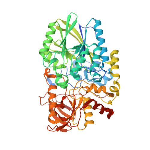
    SoxB is an essential component of the bacterial Sox sulfur oxidation pathway. SoxB contains a di-manganese(II) site and is proposed to catalyze the release of sulfate from a protein-bound cysteine S-thiosulfonate. A direct assay for SoxB activity is described. The structure of recombinant Thermus thermophilus SoxB was determined by x-ray crystallography to a resolution of 1.5 A. Structures were also determined for SoxB in complex with the substrate analogue thiosulfate and in complex with the product sulfate. A mechanistic model for SoxB is proposed based on these structures.


  • Organizational Affiliation
    • Department of Biochemistry, University of Oxford, Oxford OX1 3QU, United Kingdom.

Macromolecule Content 

  • Total Structure Weight: 64 kDa 
  • Atom Count: 5,085 
  • Modeled Residue Count: 542 
  • Deposited Residue Count: 562 
  • Unique protein chains: 1

Macromolecules

Find similar proteins by:|  3D Structure
Entity ID: 1
MoleculeChains  Sequence LengthOrganismDetailsImage
SULFUR OXIDATION PROTEIN SOXB562Thermus thermophilus HB27Mutation(s): 0 
UniProt
Find proteins for Q72IT0 (Thermus thermophilus (strain ATCC BAA-163 / DSM 7039 / HB27))
Explore Q72IT0 
Go to UniProtKB:  Q72IT0
Entity Groups
Sequence Clusters30% Identity50% Identity70% Identity90% Identity95% Identity100% Identity
UniProt GroupQ72IT0
Sequence Annotations
Expand
Reference Sequence

Small Molecules

Ligands 4 Unique
IDChains Name / Formula / InChI Key2D Diagram3D Interactions
GOL

Query on GOL


Download:Ideal Coordinates CCD File
B [auth A],
C [auth A],
D [auth A],
E [auth A],
F [auth A]
GLYCEROL
C3 H8 O3
PEDCQBHIVMGVHV-UHFFFAOYSA-N
TBU

Query on TBU


Download:Ideal Coordinates CCD File
K [auth A]
L [auth A]
M [auth A]
N [auth A]
O [auth A]
K [auth A],
L [auth A],
M [auth A],
N [auth A],
O [auth A],
P [auth A]
TERTIARY-BUTYL ALCOHOL
C4 H10 O
DKGAVHZHDRPRBM-UHFFFAOYSA-N
ACT

Query on ACT


Download:Ideal Coordinates CCD File
G [auth A],
H [auth A],
I [auth A],
J [auth A]
ACETATE ION
C2 H3 O2
QTBSBXVTEAMEQO-UHFFFAOYSA-M
MN

Query on MN


Download:Ideal Coordinates CCD File
Q [auth A],
R [auth A]
MANGANESE (II) ION
Mn
WAEMQWOKJMHJLA-UHFFFAOYSA-N

Experimental Data & Validation

Experimental Data

  • Method: X-RAY DIFFRACTION
  • Resolution: 1.50 Å
  • R-Value Free:  0.193 (Depositor), 0.186 (DCC) 
  • R-Value Work:  0.165 (Depositor), 0.163 (DCC) 
  • R-Value Observed: 0.167 (Depositor) 
Space Group: P 21 21 21
Unit Cell:
Length ( Å )Angle ( ˚ )
a = 71.345α = 90
b = 86.456β = 90
c = 95.974γ = 90
Software Package:
Software NamePurpose
BUSTER-TNTrefinement
XPREPdata reduction
SCALAdata scaling
SHELXDphasing
SHARPphasing
SOLOMONphasing

Structure Validation

View Full Validation Report


Entry History 

Deposition Data

Revision History  (Full details and data files)

  • Version 1.0: 2009-06-16
    Type: Initial release
  • Version 1.1: 2015-08-19
    Changes: Atomic model, Derived calculations, Non-polymer description, Other, Version format compliance
  • Version 1.2: 2017-07-05
    Changes: Data collection
  • Version 1.3: 2024-05-08
    Changes: Data collection, Database references, Derived calculations, Other