2WCI | pdb_00002wci

Structure of E. coli monothiol glutaredoxin GRX4 homodimer


Experimental Data Snapshot

  • Method: X-RAY DIFFRACTION
  • Resolution: 1.90 Å
  • R-Value Free: 
    0.234 (Depositor), 0.233 (DCC) 
  • R-Value Work: 
    0.183 (Depositor), 0.185 (DCC) 
  • R-Value Observed: 
    0.186 (Depositor) 

wwPDB Validation 3D Report Full Report

Validation slider image for 2WCI

Ligand Structure Quality Assessment 


This is version 1.2 of the entry. See complete history

Literature

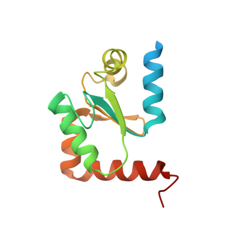
Structural Basis for Delivery of the Intact [Fe2S2] Cluster by Monothiol Glutaredoxin.

Iwema, T.Picciocchi, A.Traore, D.A.K.Ferrer, J.-L.Chauvat, F.Jacquamet, L.

(2009) Biochemistry 48: 6041

  • DOI: https://doi.org/10.1021/bi900440m
  • Primary Citation Related Structures: 
    2WCI

  • PubMed Abstract: 

    Glutaredoxins (GRX) are redox proteins which use glutathione as a cofactor and are divided into two classes, monothiol and dithiol. In each class, several GRX have been shown to form [Fe2S2] cluster coordinating homodimers. The dithiol GRX homodimer is proposed to serve as a sequestration form and its iron-sulfur cluster as an oxidative stress sensor. In contrast, the monothiol GRX homodimer has been suggested to act as a scaffold for [Fe2S2] cluster delivery. We present here the structure of a monothiol GRX homodimer (Escherichia coli GRX4) coordinating a [Fe2S2] cluster that reveals the structural basis of intact iron-sulfur cluster delivery.


  • Organizational Affiliation
    • Laboratoire de Cristallographie et Cristallogenèse des Protéines, Institut de Biologie Structurale Jean-Pierre Ebel, 5075 CEA, CNRS, Universite Joseph Fourier, 41 rue Jules Horowitz, F-38027 Grenoble, France. thomas.iwema@ibs.fr

Macromolecule Content 

  • Total Structure Weight: 30.94 kDa 
  • Atom Count: 1,993 
  • Modeled Residue Count: 225 
  • Deposited Residue Count: 270 
  • Unique protein chains: 1

Macromolecules

Find similar proteins by:|  3D Structure
Entity ID: 1
MoleculeChains  Sequence LengthOrganismDetailsImage
GLUTAREDOXIN-4
A, B
135Escherichia coliMutation(s): 0 
UniProt
Find proteins for P0AC69 (Escherichia coli (strain K12))
Explore P0AC69 
Go to UniProtKB:  P0AC69
Entity Groups
Sequence Clusters30% Identity50% Identity70% Identity90% Identity95% Identity100% Identity
UniProt GroupP0AC69
Sequence Annotations
Expand
Reference Sequence

Biologically Interesting Molecules (External Reference) 

1 Unique

Experimental Data & Validation

Experimental Data

  • Method: X-RAY DIFFRACTION
  • Resolution: 1.90 Å
  • R-Value Free:  0.234 (Depositor), 0.233 (DCC) 
  • R-Value Work:  0.183 (Depositor), 0.185 (DCC) 
  • R-Value Observed: 0.186 (Depositor) 
Space Group: P 43 21 2
Unit Cell:
Length ( Å )Angle ( ˚ )
a = 94.3α = 90
b = 94.3β = 90
c = 62.66γ = 90
Software Package:
Software NamePurpose
REFMACrefinement
XDSdata reduction
XSCALEdata scaling
SHELXphasing

Structure Validation

View Full Validation Report


Ligand Structure Quality Assessment 


Entry History 

Deposition Data

Revision History  (Full details and data files)

  • Version 1.0: 2009-06-23
    Type: Initial release
  • Version 1.1: 2012-02-29
    Changes: Advisory, Atomic model, Database references, Derived calculations, Non-polymer description, Other, Structure summary
  • Version 1.2: 2024-05-08
    Changes: Data collection, Database references, Derived calculations, Other