2WAM | pdb_00002wam

Crystal structure of Mycobacterium tuberculosis unknown function protein Rv2714


Experimental Data Snapshot

  • Method: X-RAY DIFFRACTION
  • Resolution: 2.60 Å
  • R-Value Free: 
    0.221 (Depositor), 0.220 (DCC) 
  • R-Value Work: 
    0.175 (Depositor), 0.173 (DCC) 
  • R-Value Observed: 
    0.177 (Depositor) 

Starting Model: other
View more details

wwPDB Validation 3D Report Full Report

Validation slider image for 2WAM

This is version 1.3 of the entry. See complete history

Literature

Structure of Mycobacterium Tuberculosis Rv2714, a Representative of a Duplicated Gene Family in Actinobacteria.

Grana, M.Bellinzoni, M.Miras, I.Fiex-Vandal, C.Haouz, A.Shepard, W.Buschiazzo, A.Alzari, P.M.

(2009) Acta Crystallogr Sect F Struct Biol Cryst Commun 65: 972

  • DOI: https://doi.org/10.1107/S1744309109035027
  • Primary Citation Related Structures: 
    2WAM

  • PubMed Abstract: 

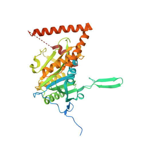
    The gene Rv2714 from Mycobacterium tuberculosis, which codes for a hypothetical protein of unknown function, is a representative member of a gene family that is largely confined to the order Actinomycetales of Actinobacteria. Sequence analysis indicates the presence of two paralogous genes in most mycobacterial genomes and suggests that gene duplication was an ancient event in bacterial evolution. The crystal structure of Rv2714 has been determined at 2.6 A resolution, revealing a trimer in which the topology of the protomer core is similar to that observed in a functionally diverse set of enzymes, including purine nucleoside phosphorylases, some carboxypeptidases, bacterial peptidyl-tRNA hydrolases and even the plastidic form of an intron splicing factor. However, some structural elements, such as a beta-hairpin insertion involved in protein oligomerization and a C-terminal alpha-helical domain that serves as a lid to the putative substrate-binding (or ligand-binding) site, are only found in Rv2714 bacterial homologues and represent specific signatures of this protein family.


  • Organizational Affiliation
    • Institut Pasteur, Unité de Biochimie Structurale, URA CNRS 2185, 25 Rue du Dr Roux, 75724 Paris, France.

Macromolecule Content 

  • Total Structure Weight: 116.64 kDa 
  • Atom Count: 6,825 
  • Modeled Residue Count: 856 
  • Deposited Residue Count: 1,053 
  • Unique protein chains: 1

Macromolecules

Find similar proteins by:|  3D Structure
Entity ID: 1
MoleculeChains  Sequence LengthOrganismDetailsImage
CONSERVED HYPOTHETICAL ALANINE AND LEUCINE RICH PROTEIN
A, B, C
351Mycobacterium tuberculosis H37RvMutation(s): 0 
UniProt
Find proteins for O07213 (Mycobacterium tuberculosis (strain CDC 1551 / Oshkosh))
Explore O07213 
Go to UniProtKB:  O07213
Entity Groups
Sequence Clusters30% Identity50% Identity70% Identity90% Identity95% Identity100% Identity
UniProt GroupO07213
Sequence Annotations
Expand
Reference Sequence

Experimental Data & Validation

Experimental Data

  • Method: X-RAY DIFFRACTION
  • Resolution: 2.60 Å
  • R-Value Free:  0.221 (Depositor), 0.220 (DCC) 
  • R-Value Work:  0.175 (Depositor), 0.173 (DCC) 
  • R-Value Observed: 0.177 (Depositor) 
Space Group: P 32 2 1
Unit Cell:
Length ( Å )Angle ( ˚ )
a = 140.493α = 90
b = 140.493β = 90
c = 129.77γ = 120
Software Package:
Software NamePurpose
PHENIXrefinement
XDSdata reduction
SCALAdata scaling
PHASERphasing

Structure Validation

View Full Validation Report


Entry History 

Deposition Data

Revision History  (Full details and data files)

  • Version 1.0: 2009-10-06
    Type: Initial release
  • Version 1.1: 2011-05-08
    Changes: Version format compliance
  • Version 1.2: 2011-07-13
    Changes: Version format compliance
  • Version 1.3: 2024-05-01
    Changes: Data collection, Database references, Derived calculations, Other, Refinement description