2W9O | pdb_00002w9o

Solution structure of jerdostatin from Trimeresurus jerdonii


Experimental Data Snapshot

  • Method: SOLUTION NMR
  • Conformers Calculated: 50 
  • Conformers Submitted: 40 
  • Selection Criteria: LOWEST TOTAL ENERGY 

wwPDB Validation 3D Report Full Report

Validation slider image for 2W9O

This is version 1.4 of the entry. See complete history

Literature

NMR Structure and Dynamics of Recombinant Wild-Type and Mutated Jerdostatin, a Selective Inhibitor of Integrin Alpha1 Beta1

Carbajo, R.J.Sanz, L.Mosulen, S.Perez, A.Marcinkiewicz, C.Pineda-Lucena, A.Calvete, J.J.

(2011) Proteins 79: 2530

  • DOI: https://doi.org/10.1002/prot.23076
  • Primary Citation Related Structures: 
    2W9O, 2W9U, 2W9V, 2W9W

  • PubMed Abstract: 

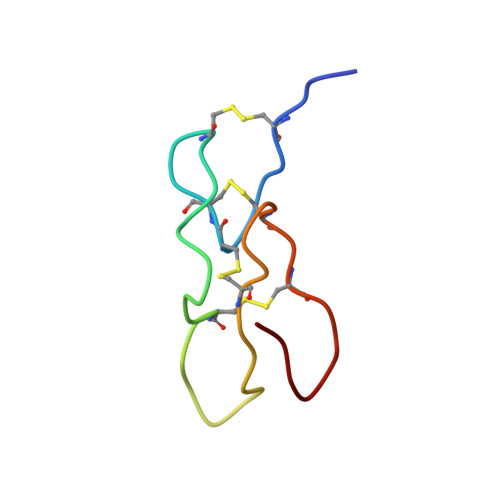
    NMR analysis of four recombinant jerdostatin molecules was assessed to define the structural basis of two naturally occurring gain-of-function events: C-terminal dipeptide processing and mutation of the active residue K21 to arginine. Removal of the highly mobile and a bulky C-terminal dipeptide produced pronounced chemical shift changes in the sequentially unconnected but spatially nearby α(1)β(1) inhibitory loop. Analysis of chemical shift divergence and (15)N backbone relaxation dynamics indicated differences in motions in the picosecond to nanosecond time scale, and the higher T(2) rate of S25, S26, and H27 of rJerK21 point to a slowdown in the microsecond to millisecond motions of these residues when compared with rJerR21. The evidence presented in this article converges on the hypothesis that dynamic differences between the α(1)β(1) recognition loops of rJerR21 and rJerK21 may influence the thermodynamics of their receptor recognition and binding. A decrease in the μs-ms time scale may impair the binding affinity by reducing the rate of possible conformations that the rJerK21 can adopt in this time scale.


  • Organizational Affiliation
    • Department of Chemical Biology, Centro de Investigación Príncipe Felipe, Valencia, Spain. rcarbajo@cipf.es

Macromolecule Content 

  • Total Structure Weight: 4.91 kDa 
  • Atom Count: 335 
  • Modeled Residue Count: 46 
  • Deposited Residue Count: 46 
  • Unique protein chains: 1

Macromolecules

Find similar proteins by:|  3D Structure
Entity ID: 1
MoleculeChains  Sequence LengthOrganismDetailsImage
SHORT DISINTEGRIN JERDOSTATIN46Protobothrops jerdoniiMutation(s): 0 
UniProt
Find proteins for Q7ZZM2 (Protobothrops jerdonii)
Explore Q7ZZM2 
Go to UniProtKB:  Q7ZZM2
Entity Groups
Sequence Clusters30% Identity50% Identity70% Identity90% Identity95% Identity100% Identity
UniProt GroupQ7ZZM2
Sequence Annotations
Expand
Reference Sequence

Experimental Data & Validation

Experimental Data

  • Method: SOLUTION NMR
  • Conformers Calculated: 50 
  • Conformers Submitted: 40 
  • Selection Criteria: LOWEST TOTAL ENERGY 

Structure Validation

View Full Validation Report


Entry History 

Deposition Data

Revision History  (Full details and data files)

  • Version 1.0: 2010-03-31
    Type: Initial release
  • Version 1.1: 2011-11-09
    Changes: Database references, Version format compliance
  • Version 1.2: 2020-01-15
    Changes: Data collection, Other
  • Version 1.3: 2023-06-14
    Changes: Database references, Other
  • Version 1.4: 2024-11-20
    Changes: Data collection, Database references, Structure summary