The Structure of the Conserved Neurotrophic Factors Manf and Cdnf Explains Why They are Bifunctional.
Parkash, V., Lindholm, P., Peranen, J., Kalkkinen, N., Oksanen, E., Saarma, M., Leppanen, V.M., Goldman, A.(2009) Protein Eng Des Sel 22: 233
- PubMed: 19258449
- DOI: https://doi.org/10.1093/protein/gzn080
- Primary Citation of Related Structures:
2W50, 2W51 - PubMed Abstract:
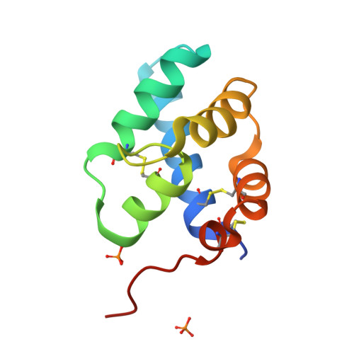
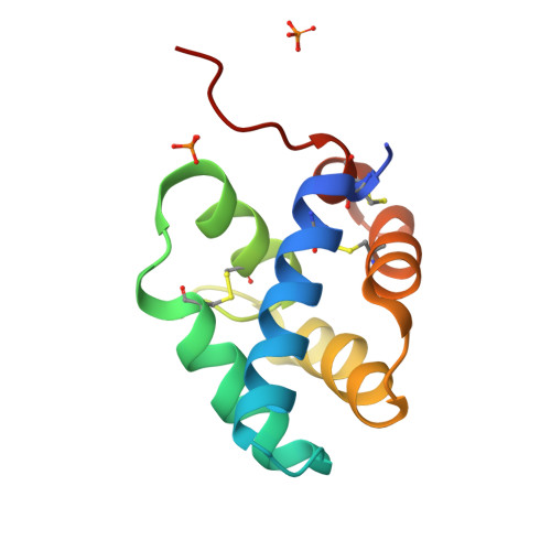
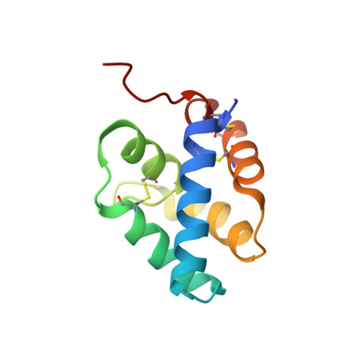
We have solved the structures of mammalian mesencephalic astrocyte-derived neurotrophic factor (MANF) and conserved dopamine neurotrophic factor (CDNF). CDNF protects and repairs midbrain dopaminergic neurons in vivo; MANF supports their survival in culture and is also cytoprotective against endoplasmic reticulum (ER) stress. Neither protein structure resembles any known growth factor but the N-terminal domain is a saposin-like lipid-binding domain. MANF and CDNF may thus bind lipids or membranes. Consistent with this, there are two patches of conserved lysines and arginines. The natively unfolded MANF C-terminus contains a CKGC disulphide bridge, such as reductases and disulphide isomerases, consistent with a role in ER stress response. The structure thus explains why MANF and CDNF are bifunctional; neurotrophic activity may reside in the N-terminal domain and ER stress response in the C-terminal domain. Finally, we identified three changes, (MANF)I10-->K(CDNF), (MANF)E79-->M(CDNF) and (MANF)K88-->L(CDNF), that may account for the biological differences between the proteins.
Organizational Affiliation:
Institute of Biotechnology, University of Helsinki, Helsinki, Finland.


