2W4U | pdb_00002w4u

Isometrically contracting insect asynchronous flight muscle quick frozen after a length step


Experimental Data Snapshot

  • Method: ELECTRON MICROSCOPY
  • Aggregation State: FILAMENT 
  • Reconstruction Method: HELICAL 

wwPDB Validation 3D Report Full Report

Validation slider image for 2W4U

This is version 1.5 of the entry. See complete history

Literature

Structural Changes in Isometrically Contracting Insect Flight Muscle Trapped Following a Mechanical Perturbation.

Wu, S.Liu, J.Reedy, M.C.Perz-Edwards, R.J.Tregear, R.T.Winkler, H.Franzini-Armstrong, C.Sasaki, H.Lucaveche, C.Goldman, Y.E.Reedy, M.K.Taylor, K.A.

(2012) PLoS One 7: 39422

  • DOI: https://doi.org/10.1371/journal.pone.0039422
  • Primary Citation Related Structures: 
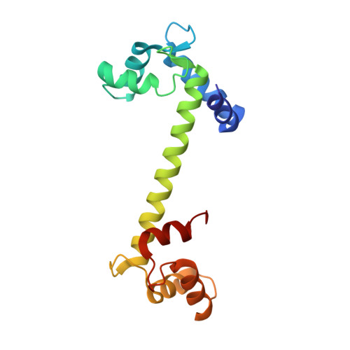
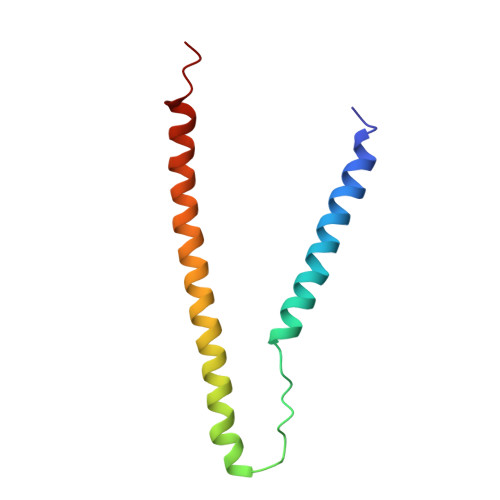
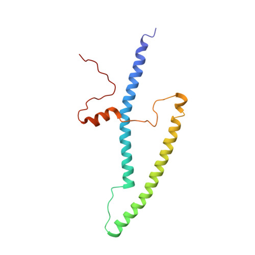
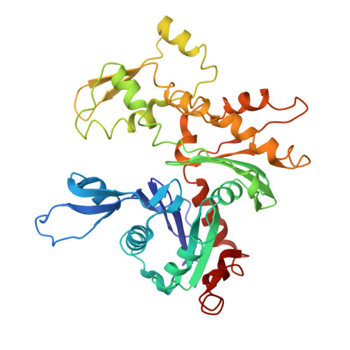
    2W4H, 2W4U, 2W4V, 2W4W

  • PubMed Abstract: 

    The application of rapidly applied length steps to actively contracting muscle is a classic method for synchronizing the response of myosin cross-bridges so that the average response of the ensemble can be measured. Alternatively, electron tomography (ET) is a technique that can report the structure of the individual members of the ensemble. We probed the structure of active myosin motors (cross-bridges) by applying 0.5% changes in length (either a stretch or a release) within 2 ms to isometrically contracting insect flight muscle (IFM) fibers followed after 5-6 ms by rapid freezing against a liquid helium cooled copper mirror. ET of freeze-substituted fibers, embedded and thin-sectioned, provides 3-D cross-bridge images, sorted by multivariate data analysis into ~40 classes, distinct in average structure, population size and lattice distribution. Individual actin subunits are resolved facilitating quasi-atomic modeling of each class average to determine its binding strength (weak or strong) to actin. ~98% of strong-binding acto-myosin attachments present after a length perturbation are confined to "target zones" of only two actin subunits located exactly midway between successive troponin complexes along each long-pitch helical repeat of actin. Significant changes in the types, distribution and structure of actin-myosin attachments occurred in a manner consistent with the mechanical transients. Most dramatic is near disappearance, after either length perturbation, of a class of weak-binding cross-bridges, attached within the target zone, that are highly likely to be precursors of strong-binding cross-bridges. These weak-binding cross-bridges were originally observed in isometrically contracting IFM. Their disappearance following a quick stretch or release can be explained by a recent kinetic model for muscle contraction, as behaviour consistent with their identification as precursors of strong-binding cross-bridges. The results provide a detailed model for contraction in IFM that may be applicable to contraction in other types of muscle.


  • Organizational Affiliation
    • Institute of Molecular Biophysics, Florida State University, Tallahassee, Florida, United States of America.

Macromolecule Content 

  • Total Structure Weight: 1,100.08 kDa 
  • Atom Count: 69,221 
  • Modeled Residue Count: 8,759 
  • Deposited Residue Count: 9,728 
  • Unique protein chains: 5

Macromolecules

Find similar proteins by:|  3D Structure
Entity ID: 1
MoleculeChains  Sequence LengthOrganismDetailsImage
TROPONIN C, SKELETAL MUSCLEA [auth 0],
D [auth 3],
G [auth 6],
J [auth 9]
159Gallus gallusMutation(s): 0 
UniProt
Find proteins for P02588 (Gallus gallus)
Explore P02588 
Go to UniProtKB:  P02588
Entity Groups
Sequence Clusters30% Identity50% Identity70% Identity90% Identity95% Identity100% Identity
UniProt GroupP02588
Sequence Annotations
Expand
Reference Sequence
Find similar proteins by:|  3D Structure
Entity ID: 2
MoleculeChains  Sequence LengthOrganismDetailsImage
TROPONIN T, FAST SKELETAL MUSCLE ISOFORMSB [auth 1],
E [auth 4],
H [auth 7],
IA [auth Y]
90Gallus gallusMutation(s): 0 
UniProt
Find proteins for P12620 (Gallus gallus)
Explore P12620 
Go to UniProtKB:  P12620
Entity Groups
Sequence Clusters30% Identity50% Identity70% Identity90% Identity95% Identity100% Identity
UniProt GroupP12620
Sequence Annotations
Expand
Reference Sequence
Find similar proteins by:|  3D Structure
Entity ID: 3
MoleculeChains  Sequence LengthOrganismDetailsImage
TROPONIN I, FAST SKELETAL MUSCLEC [auth 2],
F [auth 5],
I [auth 8],
JA [auth Z]
141Gallus gallusMutation(s): 0 
UniProt
Find proteins for P68246 (Gallus gallus)
Explore P68246 
Go to UniProtKB:  P68246
Entity Groups
Sequence Clusters30% Identity50% Identity70% Identity90% Identity95% Identity100% Identity
UniProt GroupP68246
Sequence Annotations
Expand
Reference Sequence
Find similar proteins by:|  3D Structure
Entity ID: 4
MoleculeChains  Sequence LengthOrganismDetailsImage
TROPOMYOSIN ALPHA-1 CHAIN277Oryctolagus cuniculusMutation(s): 0 
UniProt
Find proteins for P58772 (Oryctolagus cuniculus)
Explore P58772 
Go to UniProtKB:  P58772
Entity Groups
Sequence Clusters30% Identity50% Identity70% Identity90% Identity95% Identity100% Identity
UniProt GroupP58772
Sequence Annotations
Expand
Reference Sequence
Find similar proteins by:|  3D Structure
Entity ID: 5
MoleculeChains  Sequence LengthOrganismDetailsImage
ACTIN, ALPHA SKELETAL MUSCLE372Oryctolagus cuniculusMutation(s): 0 
EC: 3.6.4
UniProt
Find proteins for P68135 (Oryctolagus cuniculus)
Explore P68135 
Go to UniProtKB:  P68135
Entity Groups
Sequence Clusters30% Identity50% Identity70% Identity90% Identity95% Identity100% Identity
UniProt GroupP68135
Sequence Annotations
Expand
Reference Sequence

Experimental Data & Validation

Experimental Data

  • Method: ELECTRON MICROSCOPY
  • Aggregation State: FILAMENT 
  • Reconstruction Method: HELICAL 

Structure Validation

View Full Validation Report


Entry History 

Deposition Data

Revision History  (Full details and data files)

  • Version 1.0: 2010-08-25
    Type: Initial release
  • Version 1.1: 2012-07-18
    Changes: Database references
  • Version 1.2: 2012-08-08
    Changes: Database references, Version format compliance
  • Version 1.3: 2017-04-19
    Changes: Other
  • Version 1.4: 2019-10-23
    Changes: Author supporting evidence, Data collection, Other
  • Version 1.5: 2024-05-08
    Changes: Data collection, Database references