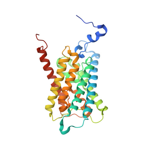
2W1P

1.4 Angstrom crystal structure of P.pastoris aquaporin, Aqy1, in a closed conformation at pH 8.0


Experimental Data Snapshot

  • Method: X-RAY DIFFRACTION
  • Resolution: 1.40 Å
  • R-Value Free: 0.178 
  • R-Value Work: 0.160 
  • R-Value Observed: 0.161 

Starting Model: experimental
View more details

wwPDB Validation   3D Report Full Report


Ligand Structure Quality Assessment 


This is version 1.3 of the entry. See complete history


Literature

Crystal Structure of a Yeast Aquaporin at 1.15 A Reveals a Novel Gating Mechanism

Fischer, G.Kosinska-Eriksson, U.Aponte-Santamaria, C.Palmgren, M.Geijer, C.Hedfalk, K.Hohmann, S.De Groot, B.L.Neutze, R.Lindkvist-Petersson, K.

(2009) PLoS Biol 7: 130

  • DOI: https://doi.org/10.1371/journal.pbio.1000130
  • Primary Citation of Related Structures:  
    2W1P, 2W2E

  • PubMed Abstract: 

    Aquaporins are transmembrane proteins that facilitate the flow of water through cellular membranes. An unusual characteristic of yeast aquaporins is that they frequently contain an extended N terminus of unknown function. Here we present the X-ray structure of the yeast aquaporin Aqy1 from Pichia pastoris at 1.15 A resolution. Our crystal structure reveals that the water channel is closed by the N terminus, which arranges as a tightly wound helical bundle, with Tyr31 forming H-bond interactions to a water molecule within the pore and thereby occluding the channel entrance. Nevertheless, functional assays show that Aqy1 has appreciable water transport activity that aids survival during rapid freezing of P. pastoris. These findings establish that Aqy1 is a gated water channel. Mutational studies in combination with molecular dynamics simulations imply that gating may be regulated by a combination of phosphorylation and mechanosensitivity.


  • Organizational Affiliation

    Department of Chemistry, Biochemistry and Biophysics, University of Gothenburg, Göteborg, Sweden.


Macromolecules
Find similar proteins by:  (by identity cutoff)  |  3D Structure
Entity ID: 1
MoleculeChains Sequence LengthOrganismDetailsImage
AQUAPORIN PIP2-7 7;279Komagataella pastorisMutation(s): 0 
Membrane Entity: Yes 
UniProt
Find proteins for F2QVG4 (Komagataella phaffii (strain ATCC 76273 / CBS 7435 / CECT 11047 / NRRL Y-11430 / Wegner 21-1))
Explore F2QVG4 
Go to UniProtKB:  F2QVG4
Entity Groups  
Sequence Clusters30% Identity50% Identity70% Identity90% Identity95% Identity100% Identity
UniProt GroupF2QVG4
Sequence Annotations
Expand
  • Reference Sequence
Experimental Data & Validation

Experimental Data

  • Method: X-RAY DIFFRACTION
  • Resolution: 1.40 Å
  • R-Value Free: 0.178 
  • R-Value Work: 0.160 
  • R-Value Observed: 0.161 
  • Space Group: I 4
Unit Cell:
Length ( Å )Angle ( ˚ )
a = 90.929α = 90
b = 90.929β = 90
c = 80.624γ = 90
Software Package:
Software NamePurpose
REFMACrefinement
MOSFLMdata reduction
SCALAdata scaling
PHASERphasing

Structure Validation

View Full Validation Report


Ligand Structure Quality Assessment 


Entry History 

Revision History  (Full details and data files)

  • Version 1.0: 2009-06-16
    Type: Initial release
  • Version 1.1: 2012-04-11
    Changes: Database references, Derived calculations, Non-polymer description, Other, Refinement description, Source and taxonomy, Structure summary, Version format compliance
  • Version 1.2: 2020-07-29
    Type: Remediation
    Reason: Carbohydrate remediation
    Changes: Advisory, Data collection, Derived calculations, Other, Structure summary
  • Version 1.3: 2023-12-13
    Changes: Advisory, Data collection, Database references, Refinement description, Structure summary