2W1A | pdb_00002w1a

Non-covalent complex between dahp synthase and chorismate mutase from Mycobacterium tuberculosis with bound tsa


Experimental Data Snapshot

  • Method: X-RAY DIFFRACTION
  • Resolution: 2.35 Å
  • R-Value Free: 
    0.229 (Depositor), 0.243 (DCC) 
  • R-Value Work: 
    0.191 (Depositor), 0.208 (DCC) 
  • R-Value Observed: 
    0.193 (Depositor) 

wwPDB Validation 3D Report Full Report

Validation slider image for 2W1A

Ligand Structure Quality Assessment 


This is version 1.2 of the entry. See complete history

Literature

Structure and Function of a Complex between Chorismate Mutase and Dahp Synthase: Efficiency Boost for the Junior Partner.

Sasso, S.Okvist, M.Roderer, K.Gamper, M.Codoni, G.Krengel, U.Kast, P.

(2009) EMBO J 28: 2128

  • DOI: https://doi.org/10.1038/emboj.2009.165
  • Primary Citation Related Structures: 
    2VKL, 2W19, 2W1A

  • PubMed Abstract: 

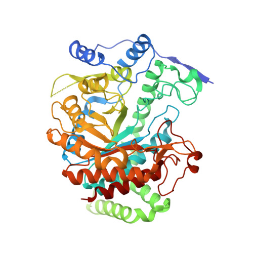
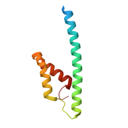
    Chorismate mutase catalyzes a key step in the shikimate biosynthetic pathway towards phenylalanine and tyrosine. Curiously, the intracellular chorismate mutase of Mycobacterium tuberculosis (MtCM; Rv0948c) has poor activity and lacks prominent active-site residues. However, its catalytic efficiency increases >100-fold on addition of DAHP synthase (MtDS; Rv2178c), another shikimate-pathway enzyme. The 2.35 A crystal structure of the MtCM-MtDS complex bound to a transition-state analogue shows a central core formed by four MtDS subunits sandwiched between two MtCM dimers. Structural comparisons imply catalytic activation to be a consequence of the repositioning of MtCM active-site residues on binding to MtDS. The mutagenesis of the C-terminal extrusion of MtCM establishes conserved residues as part of the activation machinery. The chorismate-mutase activity of the complex, but not of MtCM alone, is inhibited synergistically by phenylalanine and tyrosine. The complex formation thus endows the shikimate pathway of M. tuberculosis with an important regulatory feature. Experimental evidence suggests that such non-covalent enzyme complexes comprising an AroQ(delta) subclass chorismate mutase like MtCM are abundant in the bacterial order Actinomycetales.


  • Organizational Affiliation
    • Laboratory of Organic Chemistry, ETH Zurich, CH-8093 Zurich, Switzerland.

Macromolecule Content 

  • Total Structure Weight: 125.86 kDa 
  • Atom Count: 8,446 
  • Modeled Residue Count: 1,057 
  • Deposited Residue Count: 1,124 
  • Unique protein chains: 2

Macromolecules

Find similar proteins by:|  3D Structure
Entity ID: 1
MoleculeChains  Sequence LengthOrganismDetailsImage
3-DEOXY-D-ARABINO-HEPTULOSONATE 7-PHOSPHATE SYNTHASE AROG
A, B
472Mycobacterium tuberculosis H37RvMutation(s): 0 
EC: 2.5.1.54
UniProt
Find proteins for O53512 (Mycobacterium tuberculosis (strain ATCC 25618 / H37Rv))
Explore O53512 
Go to UniProtKB:  O53512
Entity Groups
Sequence Clusters30% Identity50% Identity70% Identity90% Identity95% Identity100% Identity
UniProt GroupO53512
Sequence Annotations
Expand
Reference Sequence
Find similar proteins by:|  3D Structure
Entity ID: 2
MoleculeChains  Sequence LengthOrganismDetailsImage
CHORISMATE MUTASE
C, D
90Mycobacterium tuberculosis H37RvMutation(s): 0 
EC: 5.4.99.5
UniProt
Find proteins for P9WIC1 (Mycobacterium tuberculosis (strain ATCC 25618 / H37Rv))
Explore P9WIC1 
Go to UniProtKB:  P9WIC1
Entity Groups
Sequence Clusters30% Identity50% Identity70% Identity90% Identity95% Identity100% Identity
UniProt GroupP9WIC1
Sequence Annotations
Expand
Reference Sequence

Small Molecules

Ligands 6 Unique
IDChains Name / Formula / InChI Key2D Diagram3D Interactions
CE1

Query on CE1


Download:Ideal Coordinates CCD File
G [auth A]O-DODECANYL OCTAETHYLENE GLYCOL
C28 H58 O9
YYELLDKEOUKVIQ-UHFFFAOYSA-N
TSA

Query on TSA


Download:Ideal Coordinates CCD File
O [auth C],
P [auth D]
8-HYDROXY-2-OXA-BICYCLO[3.3.1]NON-6-ENE-3,5-DICARBOXYLIC ACID
C10 H12 O6
KRZHNRULRHECRF-JQCUSGDOSA-N
PG4

Query on PG4


Download:Ideal Coordinates CCD File
H [auth A]TETRAETHYLENE GLYCOL
C8 H18 O5
UWHCKJMYHZGTIT-UHFFFAOYSA-N
SO4

Query on SO4


Download:Ideal Coordinates CCD File
F [auth A],
J [auth B],
K [auth B]
SULFATE ION
O4 S
QAOWNCQODCNURD-UHFFFAOYSA-L
GOL

Query on GOL


Download:Ideal Coordinates CCD File
I [auth A],
M [auth B],
N [auth B]
GLYCEROL
C3 H8 O3
PEDCQBHIVMGVHV-UHFFFAOYSA-N
MN

Query on MN


Download:Ideal Coordinates CCD File
E [auth A],
L [auth B]
MANGANESE (II) ION
Mn
WAEMQWOKJMHJLA-UHFFFAOYSA-N

Experimental Data & Validation

Experimental Data

  • Method: X-RAY DIFFRACTION
  • Resolution: 2.35 Å
  • R-Value Free:  0.229 (Depositor), 0.243 (DCC) 
  • R-Value Work:  0.191 (Depositor), 0.208 (DCC) 
  • R-Value Observed: 0.193 (Depositor) 
Space Group: P 32 2 1
Unit Cell:
Length ( Å )Angle ( ˚ )
a = 205.91α = 90
b = 205.91β = 90
c = 67.24γ = 120
Software Package:
Software NamePurpose
REFMACrefinement
MOSFLMdata reduction
SCALAdata scaling

Structure Validation

View Full Validation Report


Ligand Structure Quality Assessment 


Entry History 

Deposition Data

Revision History  (Full details and data files)

  • Version 1.0: 2009-07-07
    Type: Initial release
  • Version 1.1: 2012-04-11
    Changes: Atomic model, Derived calculations, Non-polymer description, Other, Structure summary, Version format compliance
  • Version 1.2: 2024-05-08
    Changes: Data collection, Database references, Derived calculations, Other