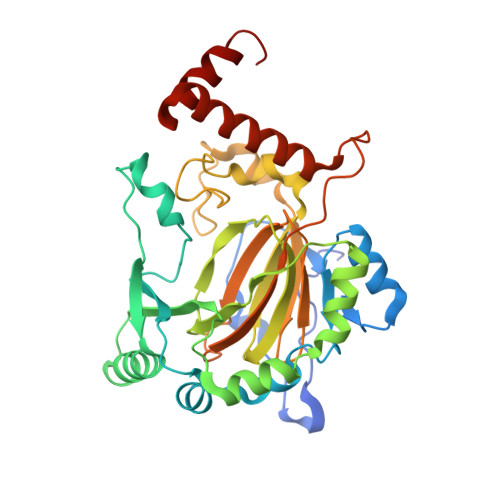
2W0X

FACTOR INHIBITING HIF-1 ALPHA WITH PYRIDINE 2,4 DICARBOXYLIC ACID


Experimental Data Snapshot

  • Method: X-RAY DIFFRACTION
  • Resolution: 2.12 Å
  • R-Value Free: 0.262 
  • R-Value Work: 0.210 
  • R-Value Observed: 0.213 

Starting Model: experimental
View more details

wwPDB Validation   3D Report Full Report


Ligand Structure Quality Assessment 


This is version 1.5 of the entry. See complete history


Literature

Structural basis for binding of cyclic 2-oxoglutarate analogues to factor-inhibiting hypoxia-inducible factor.

Conejo-Garcia, A.McDonough, M.A.Loenarz, C.McNeill, L.A.Hewitson, K.S.Ge, W.Lienard, B.M.Schofield, C.J.Clifton, I.J.

(2010) Bioorg Med Chem Lett 20: 6125-6128

  • DOI: https://doi.org/10.1016/j.bmcl.2010.08.032
  • Primary Citation of Related Structures:  
    2W0X, 2WA3, 2WA4

  • PubMed Abstract: 

    Aromatic analogues of the 2-oxoglutarate co-substrate of the hypoxia-inducible factor hydroxylases are shown to bind at the active site iron: Pyridine-2,4-dicarboxylate binds as anticipated with a single molecule chelating the iron in a bidentate manner. The binding mode of a hydroxamic acid analogue, at least in the crystalline state, is unusual because two molecules of the inhibitor are observed at the active site and partial displacement of the iron binding aspartyl residue was observed.


  • Organizational Affiliation

    The Department of Chemistry, Chemistry Research Laboratory, University of Oxford, Oxford, United Kingdom.


Macromolecules
Find similar proteins by:  (by identity cutoff)  |  3D Structure
Entity ID: 1
MoleculeChains Sequence LengthOrganismDetailsImage
HYPOXIA-INDUCIBLE FACTOR 1 ALPHA INHIBITOR349Homo sapiensMutation(s): 0 
EC: 1.14.11.16 (PDB Primary Data), 1.14.11.30 (UniProt)
UniProt & NIH Common Fund Data Resources
Find proteins for Q9NWT6 (Homo sapiens)
Explore Q9NWT6 
Go to UniProtKB:  Q9NWT6
PHAROS:  Q9NWT6
GTEx:  ENSG00000166135 
Entity Groups  
Sequence Clusters30% Identity50% Identity70% Identity90% Identity95% Identity100% Identity
UniProt GroupQ9NWT6
Sequence Annotations
Expand
  • Reference Sequence
Small Molecules
Binding Affinity Annotations 
IDSourceBinding Affinity
PD2 BindingDB:  2W0X IC50: min: 1100, max: 8600 (nM) from 2 assay(s)
PDBBind:  2W0X IC50: 6.25e+5 (nM) from 1 assay(s)
Experimental Data & Validation

Experimental Data

  • Method: X-RAY DIFFRACTION
  • Resolution: 2.12 Å
  • R-Value Free: 0.262 
  • R-Value Work: 0.210 
  • R-Value Observed: 0.213 
  • Space Group: P 41 21 2
Unit Cell:
Length ( Å )Angle ( ˚ )
a = 86.471α = 90
b = 86.471β = 90
c = 147.237γ = 90
Software Package:
Software NamePurpose
PHENIXrefinement
MOSFLMdata reduction
SCALAdata scaling

Structure Validation

View Full Validation Report


Ligand Structure Quality Assessment 


Entry History 

Deposition Data

Revision History  (Full details and data files)

  • Version 1.0: 2009-11-24
    Type: Initial release
  • Version 1.1: 2011-05-08
    Changes: Version format compliance
  • Version 1.2: 2011-07-13
    Changes: Version format compliance
  • Version 1.3: 2017-07-05
    Changes: Refinement description
  • Version 1.4: 2018-02-21
    Changes: Database references, Structure summary
  • Version 1.5: 2023-12-13
    Changes: Data collection, Database references, Derived calculations, Other, Refinement description