2W0B | pdb_00002w0b

CYP51 OF M. TUBERCULOSIS BOUND TO AN INHIBITOR 3-{[(4-METHYLPHENYL)SULFONYL]AMINO}PROPYL PYRIDIN-4-YLCARBAMATE


Experimental Data Snapshot

  • Method: X-RAY DIFFRACTION
  • Resolution: 1.56 Å
  • R-Value Free: 
    0.201 (Depositor), 0.201 (DCC) 
  • R-Value Work: 
    0.164 (Depositor), 0.163 (DCC) 
  • R-Value Observed: 
    0.168 (Depositor) 

Starting Model: experimental
View more details

wwPDB Validation 3D Report Full Report

Validation slider image for 2W0B

Ligand Structure Quality Assessment 


This is version 1.5 of the entry. See complete history

Literature

Trypanosoma cruzi CYP51 inhibitor derived from a Mycobacterium tuberculosis screen hit.

Chen, C.K.Doyle, P.S.Yermalitskaya, L.V.Mackey, Z.B.Ang, K.K.McKerrow, J.H.Podust, L.M.

(2009) PLoS Negl Trop Dis 3: e372-e372

  • DOI: https://doi.org/10.1371/journal.pntd.0000372
  • Primary Citation Related Structures: 
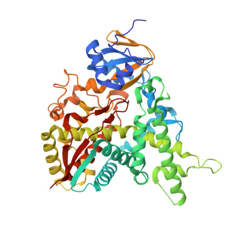
    2W09, 2W0A, 2W0B

  • PubMed Abstract: 

    The two front-line drugs for chronic Trypanosoma cruzi infections are limited by adverse side-effects and declining efficacy. One potential new target for Chagas' disease chemotherapy is sterol 14alpha-demethylase (CYP51), a cytochrome P450 enzyme involved in biosynthesis of membrane sterols. In a screening effort targeting Mycobacterium tuberculosis CYP51 (CYP51(Mt)), we previously identified the N-[4-pyridyl]-formamide moiety as a building block capable of delivering a variety of chemotypes into the CYP51 active site. In that work, the binding modes of several second generation compounds carrying this scaffold were determined by high-resolution co-crystal structures with CYP51(Mt). Subsequent assays against the CYP51 orthologue in T. cruzi, CYP51(Tc), demonstrated that two of the compounds tested in the earlier effort bound tightly to this enzyme. Both were tested in vitro for inhibitory effects against T. cruzi and the related protozoan parasite Trypanosoma brucei, the causative agent of African sleeping sickness. One of the compounds had potent, selective anti-T. cruzi activity in infected mouse macrophages. Cure of treated host cells was confirmed by prolonged incubation in the absence of the inhibiting compound. Discrimination between T. cruzi and T. brucei CYP51 by the inhibitor was largely based on the variability (phenylalanine versus isoleucine) of a single residue at a critical position in the active site. CYP51(Mt)-based crystal structure analysis revealed that the functional groups of the two tightly bound compounds are likely to occupy different spaces in the CYP51 active site, suggesting the possibility of combining the beneficial features of both inhibitors in a third generation of compounds to achieve more potent and selective inhibition of CYP51(Tc).


  • Organizational Affiliation
    • Department of Pharmaceutical Chemistry, University of California, San Francisco, California, United States of America.

Macromolecule Content 

  • Total Structure Weight: 52.44 kDa 
  • Atom Count: 3,989 
  • Modeled Residue Count: 440 
  • Deposited Residue Count: 455 
  • Unique protein chains: 1

Macromolecules

Find similar proteins by:|  3D Structure
Entity ID: 1
MoleculeChains  Sequence LengthOrganismDetailsImage
CYTOCHROME P450 51455Mycobacterium tuberculosis H37RvMutation(s): 2 
EC: 1.14.13.70 (PDB Primary Data), 1.14.15.36 (UniProt)
UniProt
Find proteins for P9WPP9 (Mycobacterium tuberculosis (strain ATCC 25618 / H37Rv))
Explore P9WPP9 
Go to UniProtKB:  P9WPP9
Entity Groups
Sequence Clusters30% Identity50% Identity70% Identity90% Identity95% Identity100% Identity
UniProt GroupP9WPP9
Sequence Annotations
Expand
Reference Sequence

Small Molecules

Ligands 2 Unique
IDChains Name / Formula / InChI Key2D Diagram3D Interactions
HEM

Query on HEM


Download:Ideal Coordinates CCD File
B [auth A]PROTOPORPHYRIN IX CONTAINING FE
C34 H32 Fe N4 O4
KABFMIBPWCXCRK-RGGAHWMASA-L
CMW

Query on CMW


Download:Ideal Coordinates CCD File
C [auth A]3-{[(4-methylphenyl)sulfonyl]amino}propyl pyridin-4-ylcarbamate
C16 H19 N3 O4 S
ITYCDQJBLCTIID-UHFFFAOYSA-N

Experimental Data & Validation

Experimental Data

  • Method: X-RAY DIFFRACTION
  • Resolution: 1.56 Å
  • R-Value Free:  0.201 (Depositor), 0.201 (DCC) 
  • R-Value Work:  0.164 (Depositor), 0.163 (DCC) 
  • R-Value Observed: 0.168 (Depositor) 
Space Group: P 21 21 21
Unit Cell:
Length ( Å )Angle ( ˚ )
a = 46.71α = 90
b = 84.813β = 90
c = 110.255γ = 90
Software Package:
Software NamePurpose
REFMACrefinement
HKL-2000data reduction
HKL-2000data scaling
MOLREPphasing

Structure Validation

View Full Validation Report


Ligand Structure Quality Assessment 


Entry History 

Deposition Data

  • Released Date: 2009-01-20 
  • Deposition Author(s): Podust, L.M.

Revision History  (Full details and data files)

  • Version 1.0: 2009-01-20
    Type: Initial release
  • Version 1.1: 2011-05-08
    Changes: Version format compliance
  • Version 1.2: 2011-07-13
    Changes: Version format compliance
  • Version 1.3: 2019-05-08
    Changes: Data collection, Experimental preparation, Other
  • Version 1.4: 2019-10-09
    Changes: Data collection, Database references, Other
  • Version 1.5: 2023-12-13
    Changes: Data collection, Database references, Derived calculations, Refinement description