2VZN | pdb_00002vzn

Crystal structure of the major allergen from fire ant venom, Sol i 3


Experimental Data Snapshot

  • Method: X-RAY DIFFRACTION
  • Resolution: 3.05 Å
  • R-Value Free: 
    0.239 (Depositor), 0.243 (DCC) 
  • R-Value Work: 
    0.201 (Depositor), 0.208 (DCC) 
  • R-Value Observed: 
    0.203 (Depositor) 

Starting Model: experimental
View more details

wwPDB Validation 3D Report Full Report

Validation slider image for 2VZN

This is version 1.4 of the entry. See complete history

Literature

Crystal Structure of the Major Allergen from Fire Ant Venom, Sol I 3

Padavattan, S.Schmidt, M.Hoffman, D.R.Markovic-Housley, Z.

(2008) J Mol Biology 383: 178

  • DOI: https://doi.org/10.1016/j.jmb.2008.08.023
  • Primary Citation Related Structures: 
    2VZN

  • PubMed Abstract: 

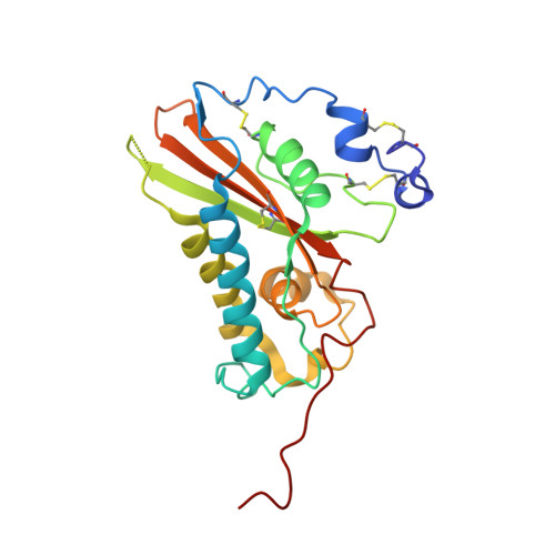
    Fire ant venom is an extremely potent allergy-inducing agent containing four major allergens, Sol i 1 to Sol i 4, which are the most frequent cause of hypersensitivity reactions to hymenoptera in the southern USA. The crystal structure of recombinant (Baculovirus) major fire ant allergen Sol i 3 has been determined to a resolution of 3.1 A by the method of molecular replacement. The secondary-structure elements of Sol i 3 are arranged in an alpha-beta-alpha sandwich fold consisting of a central antiparallel beta-sheet surrounded on both sides by alpha helices. The overall structure is very similar to that of the homologous wasp venom allergen Ves v 5 with major differences occurring in the solvent-exposed loop regions that contain amino acid insertions. Consequently, the limited conservation of surface chemical properties and topology between Sol i 3 and Ves v 5 may explain the observed lack of relevant cross-reactivity. It is concluded that Sol i 3 recognizes immunoglobulin E antibodies with a distinct set of its own epitopes, which are different from those of Ves v 5. Indeed, the molecular area in Sol i 3 covered by non-conserved residues is large enough to accommodate four unique Sol i 3 epitopes.


  • Organizational Affiliation
    • Department of Structural Biology, Biozentrum, University of Basel, CH-4056 Basel, Switzerland.

Macromolecule Content 

  • Total Structure Weight: 49.65 kDa 
  • Atom Count: 3,219 
  • Modeled Residue Count: 420 
  • Deposited Residue Count: 436 
  • Unique protein chains: 1

Macromolecules

Find similar proteins by:|  3D Structure
Entity ID: 1
MoleculeChains  Sequence LengthOrganismDetailsImage
VENOM ALLERGEN 3
A, B
218Solenopsis invictaMutation(s): 0 
UniProt
Find proteins for P35778 (Solenopsis invicta)
Explore P35778 
Go to UniProtKB:  P35778
Entity Groups
Sequence Clusters30% Identity50% Identity70% Identity90% Identity95% Identity100% Identity
UniProt GroupP35778
Sequence Annotations
Expand
Reference Sequence

Experimental Data & Validation

Experimental Data

  • Method: X-RAY DIFFRACTION
  • Resolution: 3.05 Å
  • R-Value Free:  0.239 (Depositor), 0.243 (DCC) 
  • R-Value Work:  0.201 (Depositor), 0.208 (DCC) 
  • R-Value Observed: 0.203 (Depositor) 
Space Group: P 61
Unit Cell:
Length ( Å )Angle ( ˚ )
a = 93.355α = 90
b = 93.355β = 90
c = 144.051γ = 120
Software Package:
Software NamePurpose
REFMACrefinement
MOSFLMdata reduction
SCALAdata scaling
PHASERphasing

Structure Validation

View Full Validation Report


Entry History 

Deposition Data

Revision History  (Full details and data files)

  • Version 1.0: 2008-09-09
    Type: Initial release
  • Version 1.1: 2011-07-13
    Changes: Advisory, Version format compliance
  • Version 1.2: 2019-05-15
    Changes: Data collection, Experimental preparation, Other
  • Version 1.3: 2023-12-13
    Changes: Data collection, Database references, Other, Refinement description
  • Version 1.4: 2024-11-20
    Changes: Structure summary