2VZI | pdb_00002vzi

Crystal structure of the C-terminal calponin homology domain of alpha- parvin in complex with paxillin LD4 motif


Experimental Data Snapshot

  • Method: X-RAY DIFFRACTION
  • Resolution: 2.20 Å
  • R-Value Free: 
    0.260 (Depositor), 0.251 (DCC) 
  • R-Value Work: 
    0.205 (Depositor), 0.197 (DCC) 
  • R-Value Observed: 
    0.208 (Depositor) 

Starting Model: experimental
View more details

wwPDB Validation 3D Report Full Report

Validation slider image for 2VZI

This is version 1.4 of the entry. See complete history

Literature

Structural analysis of the interactions between paxillin LD motifs and alpha-parvin.

Lorenz, S.Vakonakis, I.Lowe, E.D.Campbell, I.D.Noble, M.E.Hoellerer, M.K.

(2008) Structure 16: 1521-1531

  • DOI: https://doi.org/10.1016/j.str.2008.08.007
  • Primary Citation Related Structures: 
    2VZC, 2VZD, 2VZG, 2VZI

  • PubMed Abstract: 

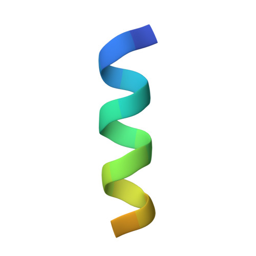
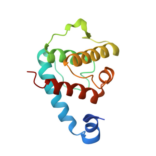
    The adaptor protein paxillin contains five conserved leucine-rich (LD) motifs that interact with a variety of focal adhesion proteins, such as alpha-parvin. Here, we report the first crystal structure of the C-terminal calponin homology domain (CH(C)) of alpha-parvin at 1.05 A resolution and show that it is able to bind all the LD motifs, with some selectivity for LD1, LD2, and LD4. Cocrystal structures with these LD motifs reveal the molecular details of their interactions with a common binding site on alpha-parvin-CH(C), which is located at the rim of the canonical fold and includes part of the inter-CH domain linker. Surprisingly, this binding site can accommodate LD motifs in two antiparallel orientations. Taken together, these results reveal an unusual degree of binding degeneracy in the paxillin/alpha-parvin system that may facilitate the assembly of dynamic signaling complexes in the cell.


  • Organizational Affiliation
    • Laboratory of Molecular Biophysics, University of Oxford, Oxford OX1 3QU, United Kingdom.

Macromolecule Content 

  • Total Structure Weight: 18.08 kDa 
  • Atom Count: 1,413 
  • Modeled Residue Count: 139 
  • Deposited Residue Count: 151 
  • Unique protein chains: 2

Macromolecules

Find similar proteins by:  Sequence   |   3D Structure  
Entity ID: 1
MoleculeChains  Sequence LengthOrganismDetailsImage
Paxillin,Paxillin20Homo sapiensMutation(s): 0 
Gene Names: PXN
UniProt & NIH Common Fund Data Resources
Find proteins for P49023 (Homo sapiens)
Explore P49023 
Go to UniProtKB:  P49023
PHAROS:  P49023
GTEx:  ENSG00000089159 
Entity Groups
Sequence Clusters30% Identity50% Identity70% Identity90% Identity95% Identity100% Identity
UniProt GroupP49023
Sequence Annotations
Expand
Reference Sequence
Find similar proteins by:|  3D Structure
Entity ID: 2
MoleculeChains  Sequence LengthOrganismDetailsImage
Alpha-parvin131Homo sapiensMutation(s): 0 
Gene Names: PARVAMXRA2
UniProt & NIH Common Fund Data Resources
Find proteins for Q9NVD7 (Homo sapiens)
Explore Q9NVD7 
Go to UniProtKB:  Q9NVD7
PHAROS:  Q9NVD7
GTEx:  ENSG00000197702 
Entity Groups
Sequence Clusters30% Identity50% Identity70% Identity90% Identity95% Identity100% Identity
UniProt GroupQ9NVD7
Sequence Annotations
Expand
Reference Sequence

Experimental Data & Validation

Experimental Data

  • Method: X-RAY DIFFRACTION
  • Resolution: 2.20 Å
  • R-Value Free:  0.260 (Depositor), 0.251 (DCC) 
  • R-Value Work:  0.205 (Depositor), 0.197 (DCC) 
  • R-Value Observed: 0.208 (Depositor) 
Space Group: C 2 2 21
Unit Cell:
Length ( Å )Angle ( ˚ )
a = 75.42α = 90
b = 94.6β = 90
c = 42.17γ = 90
Software Package:
Software NamePurpose
REFMACrefinement
MOSFLMdata reduction
SCALAdata scaling
PHASERphasing

Structure Validation

View Full Validation Report


Entry History 

Deposition Data

Revision History  (Full details and data files)

  • Version 1.0: 2008-10-28
    Type: Initial release
  • Version 1.1: 2011-07-13
    Changes: Advisory, Version format compliance
  • Version 1.2: 2012-05-30
    Changes: Other
  • Version 1.3: 2019-04-24
    Changes: Data collection, Database references, Experimental preparation, Other, Source and taxonomy, Structure summary
  • Version 1.4: 2023-12-13
    Changes: Data collection, Database references, Derived calculations, Other, Refinement description