2VXI | pdb_00002vxi

The binding of heme and zinc in Escherichia coli Bacterioferritin


Experimental Data Snapshot

  • Method: X-RAY DIFFRACTION
  • Resolution: 1.91 Å
  • R-Value Free: 
    0.216 (Depositor), 0.215 (DCC) 
  • R-Value Work: 
    0.179 (Depositor), 0.180 (DCC) 
  • R-Value Observed: 
    0.179 (Depositor) 

wwPDB Validation 3D Report Full Report

Validation slider image for 2VXI

Ligand Structure Quality Assessment 


This is version 1.3 of the entry. See complete history

Literature

The Binding of Haem and Zinc in the 1.9 A X-Ray Structure of Escherichia Coli Bacterioferritin.

Willies, S.C.Isupov, M.N.Garman, E.F.Littlechild, J.A.

(2009) J Biol Inorg Chem 14: 201

  • DOI: https://doi.org/10.1007/s00775-008-0438-8
  • Primary Citation Related Structures: 
    2VXI

  • PubMed Abstract: 

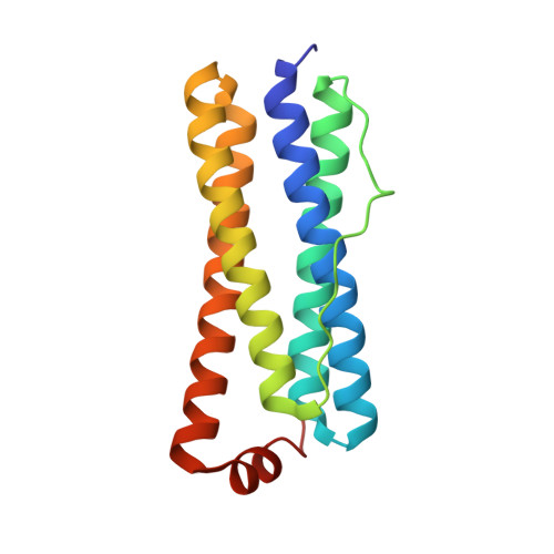
    The crystal structure of Escherichia coli bacterioferritin has been solved to 1.9 A, and shows the symmetrical binding of a haem molecule on the local twofold axis between subunits and a pair of metal atoms bound to each subunit at the ferroxidase centre. These metals have been identified as zinc by the analysis of the structure and X-ray data and confirmed by microfocused proton-induced X-ray emission experiments. For the first time the haem has been shown to be linked to both the internal and the external environments via a cluster of waters positioned above the haem molecule.


  • Organizational Affiliation
    • School of Biosciences, Henry Wellcome Building for Biocatalysis, University of Exeter, Stocker Road, Exeter, EX4 4QD, UK.

Macromolecule Content 

  • Total Structure Weight: 233.97 kDa 
  • Atom Count: 18,818 
  • Modeled Residue Count: 1,884 
  • Deposited Residue Count: 1,896 
  • Unique protein chains: 1

Macromolecules

Find similar proteins by:|  3D Structure
Entity ID: 1
MoleculeChains  Sequence LengthOrganismDetailsImage
BACTERIOFERRITIN
A, B, C, D, E
A, B, C, D, E, F, G, H, I, J, K, L
158Escherichia coli BL21Mutation(s): 0 
EC: 1.16.3.1
UniProt
Find proteins for P0ABD3 (Escherichia coli (strain K12))
Explore P0ABD3 
Go to UniProtKB:  P0ABD3
Entity Groups
Sequence Clusters30% Identity50% Identity70% Identity90% Identity95% Identity100% Identity
UniProt GroupP0ABD3
Sequence Annotations
Expand
Reference Sequence

Small Molecules

Ligands 3 Unique
IDChains Name / Formula / InChI Key2D Diagram3D Interactions
HEM

Query on HEM


Download:Ideal Coordinates CCD File
FA [auth D]
FB [auth I]
KA [auth E]
KB [auth J]
M [auth A]
FA [auth D],
FB [auth I],
KA [auth E],
KB [auth J],
M [auth A],
PB [auth K],
QA [auth F],
S [auth B],
UA [auth G],
UB [auth L],
Z [auth C],
ZA [auth H]
PROTOPORPHYRIN IX CONTAINING FE
C34 H32 Fe N4 O4
KABFMIBPWCXCRK-RGGAHWMASA-L
SO4

Query on SO4


Download:Ideal Coordinates CCD File
CA [auth C]
CB [auth H]
DA [auth C]
DB [auth H]
EA [auth C]
CA [auth C],
CB [auth H],
DA [auth C],
DB [auth H],
EA [auth C],
EB [auth H],
IA [auth D],
IB [auth I],
JA [auth D],
JB [auth I],
NA [auth E],
NB [auth J],
OA [auth E],
OB [auth J],
P [auth A],
PA [auth E],
Q [auth A],
R [auth A],
SB [auth K],
TA [auth F],
TB [auth K],
V [auth B],
W [auth B],
X [auth B],
XA [auth G],
XB [auth L],
Y [auth B],
YA [auth G],
YB [auth L]
SULFATE ION
O4 S
QAOWNCQODCNURD-UHFFFAOYSA-L
ZN

Query on ZN


Download:Ideal Coordinates CCD File
AA [auth C]
AB [auth H]
BA [auth C]
BB [auth H]
GA [auth D]
AA [auth C],
AB [auth H],
BA [auth C],
BB [auth H],
GA [auth D],
GB [auth I],
HA [auth D],
HB [auth I],
LA [auth E],
LB [auth J],
MA [auth E],
MB [auth J],
N [auth A],
O [auth A],
QB [auth K],
RA [auth F],
RB [auth K],
SA [auth F],
T [auth B],
U [auth B],
VA [auth G],
VB [auth L],
WA [auth G],
WB [auth L]
ZINC ION
Zn
PTFCDOFLOPIGGS-UHFFFAOYSA-N

Experimental Data & Validation

Experimental Data

  • Method: X-RAY DIFFRACTION
  • Resolution: 1.91 Å
  • R-Value Free:  0.216 (Depositor), 0.215 (DCC) 
  • R-Value Work:  0.179 (Depositor), 0.180 (DCC) 
  • R-Value Observed: 0.179 (Depositor) 
Space Group: P 42 21 2
Unit Cell:
Length ( Å )Angle ( ˚ )
a = 208.1α = 90
b = 208.1β = 90
c = 142.768γ = 90
Software Package:
Software NamePurpose
REFMACmodel building
SCALEPACKdata scaling
SHELXDphasing
MLPHAREphasing
DMphasing
REFMACphasing
REFMACrefinement

Structure Validation

View Full Validation Report


Ligand Structure Quality Assessment 


Entry History 

Deposition Data

Revision History  (Full details and data files)

  • Version 1.0: 2008-11-04
    Type: Initial release
  • Version 1.1: 2011-05-07
    Changes: Version format compliance
  • Version 1.2: 2011-07-13
    Changes: Version format compliance
  • Version 1.3: 2024-05-08
    Changes: Data collection, Database references, Derived calculations, Other