2VV5 | pdb_00002vv5

The open structure of MscS


Experimental Data Snapshot

  • Method: X-RAY DIFFRACTION
  • Resolution: 3.45 Å
  • R-Value Free: 
    0.312 (Depositor), 0.277 (DCC) 
  • R-Value Work: 
    0.293 (Depositor), 0.263 (DCC) 
  • R-Value Observed: 
    0.294 (Depositor) 

Starting Model: experimental
View more details

wwPDB Validation 3D Report Full Report

Validation slider image for 2VV5

This is version 1.2 of the entry. See complete history

Literature

The Structure of an Open Form of an E. Coli Mechanosensitive Channel at 3.45 A Resolution.

Wang, W.Black, S.S.Edwards, M.D.Miller, S.Morrison, E.L.Bartlett, W.Dong, C.Naismith, J.H.Booth, I.R.

(2008) Science 321: 1179

  • DOI: https://doi.org/10.1126/science.1159262
  • Primary Citation Related Structures: 
    2VV5

  • PubMed Abstract: 

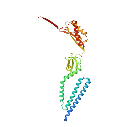
    How ion channels are gated to regulate ion flux in and out of cells is the subject of intense interest. The Escherichia coli mechanosensitive channel, MscS, opens to allow rapid ion efflux, relieving the turgor pressure that would otherwise destroy the cell. We present a 3.45 angstrom-resolution structure for the MscS channel in an open conformation. This structure has a pore diameter of approximately 13 angstroms created by substantial rotational rearrangement of the three transmembrane helices. The structure suggests a molecular mechanism that underlies MscS gating and its decay of conductivity during prolonged activation. Support for this mechanism is provided by single-channel analysis of mutants with altered gating characteristics.


  • Organizational Affiliation
    • Centre for Biomolecular Sciences, The North Haugh, University of St. Andrews, KY16 9ST, Scotland, UK.

Macromolecule Content 

  • Total Structure Weight: 216.66 kDa 
  • Atom Count: 13,692 
  • Modeled Residue Count: 1,799 
  • Deposited Residue Count: 2,002 
  • Unique protein chains: 1

Macromolecules

Find similar proteins by:|  3D Structure
Entity ID: 1
MoleculeChains  Sequence LengthOrganismDetailsImage
SMALL-CONDUCTANCE MECHANOSENSITIVE CHANNEL
A, B, C, D, E
A, B, C, D, E, F, G
286Escherichia coliMutation(s): 1 
Membrane Entity: Yes 
UniProt
Find proteins for P0C0S1 (Escherichia coli (strain K12))
Explore P0C0S1 
Go to UniProtKB:  P0C0S1
Entity Groups
Sequence Clusters30% Identity50% Identity70% Identity90% Identity95% Identity100% Identity
UniProt GroupP0C0S1
Sequence Annotations
Expand
Reference Sequence

Experimental Data & Validation

Experimental Data

  • Method: X-RAY DIFFRACTION
  • Resolution: 3.45 Å
  • R-Value Free:  0.312 (Depositor), 0.277 (DCC) 
  • R-Value Work:  0.293 (Depositor), 0.263 (DCC) 
  • R-Value Observed: 0.294 (Depositor) 
Space Group: C 1 2 1
Unit Cell:
Length ( Å )Angle ( ˚ )
a = 230.88α = 90
b = 126.62β = 90.42
c = 123.22γ = 90
Software Package:
Software NamePurpose
REFMACrefinement
XDSdata reduction
MOSFLMdata reduction
SCALAdata scaling
PHASERphasing

Structure Validation

View Full Validation Report


Entry History 

Deposition Data

Revision History  (Full details and data files)

  • Version 1.0: 2008-08-05
    Type: Initial release
  • Version 1.1: 2011-07-13
    Changes: Advisory, Version format compliance
  • Version 1.2: 2023-12-13
    Changes: Data collection, Database references, Other, Refinement description