2VUP | pdb_00002vup

Crystal structure of a type II tryparedoxin-dependant peroxidase from Trypanosoma brucei


Experimental Data Snapshot

  • Method: X-RAY DIFFRACTION
  • Resolution: 2.10 Å
  • R-Value Free: 
    0.263 (Depositor), 0.258 (DCC) 
  • R-Value Work: 
    0.205 (Depositor), 0.204 (DCC) 
  • R-Value Observed: 
    0.208 (Depositor) 

Starting Model: experimental
View more details

wwPDB Validation 3D Report Full Report

Validation slider image for 2VUP

This is version 1.3 of the entry. See complete history

Literature

Structural and Mechanistic Insights Into Type II Trypanosomatid Tryparedoxin-Dependent Peroxidases.

Alphey, M.S.Konig, J.Fairlamb, A.H.

(2008) Biochem J 414: 375

  • DOI: https://doi.org/10.1042/BJ20080889
  • Primary Citation Related Structures: 
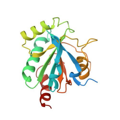
    2VUP

  • PubMed Abstract: 

    TbTDPX (Trypanosoma brucei tryparedoxin-dependent peroxidase) is a genetically validated drug target in the fight against African sleeping sickness. Despite its similarity to members of the GPX (glutathione peroxidase) family, TbTDPX2 is functional as a monomer, lacks a selenocysteine residue and relies instead on peroxidatic and resolving cysteine residues for catalysis and uses tryparedoxin rather than glutathione as electron donor. Kinetic studies indicate a saturable Ping Pong mechanism, unlike selenium-dependent GPXs, which display infinite K(m) and V(max) values. The structure of the reduced enzyme at 2.1 A (0.21 nm) resolution reveals that the catalytic thiol groups are widely separated [19 A (0.19 nm)] and thus unable to form a disulphide bond without a large conformational change in the secondary-structure architecture, as reported for certain plant GPXs. A model of the oxidized enzyme structure is presented and the implications for small-molecule inhibition are discussed.


  • Organizational Affiliation
    • Division of Biological Chemistry and Drug Discovery, College of Life Sciences, University of Dundee, Dundee DD1 5EH, Scotland, UK.

Macromolecule Content 

  • Total Structure Weight: 21.14 kDa 
  • Atom Count: 1,409 
  • Modeled Residue Count: 164 
  • Deposited Residue Count: 190 
  • Unique protein chains: 1

Macromolecules

Find similar proteins by:|  3D Structure
Entity ID: 1
MoleculeChains  Sequence LengthOrganismDetailsImage
GLUTATHIONE PEROXIDASE-LIKE PROTEIN190Trypanosoma bruceiMutation(s): 1 
EC: 1.11.1.9
UniProt
Find proteins for Q869A6 (Trypanosoma brucei)
Explore Q869A6 
Go to UniProtKB:  Q869A6
Entity Groups
Sequence Clusters30% Identity50% Identity70% Identity90% Identity95% Identity100% Identity
UniProt GroupQ869A6
Sequence Annotations
Expand
Reference Sequence

Experimental Data & Validation

Experimental Data

  • Method: X-RAY DIFFRACTION
  • Resolution: 2.10 Å
  • R-Value Free:  0.263 (Depositor), 0.258 (DCC) 
  • R-Value Work:  0.205 (Depositor), 0.204 (DCC) 
  • R-Value Observed: 0.208 (Depositor) 
Space Group: P 1 21 1
Unit Cell:
Length ( Å )Angle ( ˚ )
a = 43.26α = 90
b = 32.7β = 95.92
c = 58.31γ = 90
Software Package:
Software NamePurpose
REFMACrefinement
XDSdata reduction
XSCALEdata scaling
MOLREPphasing

Structure Validation

View Full Validation Report


Entry History 

Deposition Data

Revision History  (Full details and data files)

  • Version 1.0: 2008-06-17
    Type: Initial release
  • Version 1.1: 2011-05-08
    Changes: Version format compliance
  • Version 1.2: 2011-07-13
    Changes: Version format compliance
  • Version 1.3: 2023-12-13
    Changes: Data collection, Database references, Other, Refinement description