2VOI | pdb_00002voi

Structure of mouse A1 bound to the Bid BH3-domain


Experimental Data Snapshot

  • Method: X-RAY DIFFRACTION
  • Resolution: 2.10 Å
  • R-Value Free: 
    0.242 (Depositor), 0.249 (DCC) 
  • R-Value Work: 
    0.207 (Depositor), 0.210 (DCC) 
  • R-Value Observed: 
    0.209 (Depositor) 

Starting Model: experimental
View more details

wwPDB Validation 3D Report Full Report

Validation slider image for 2VOI

This is version 1.2 of the entry. See complete history

Literature

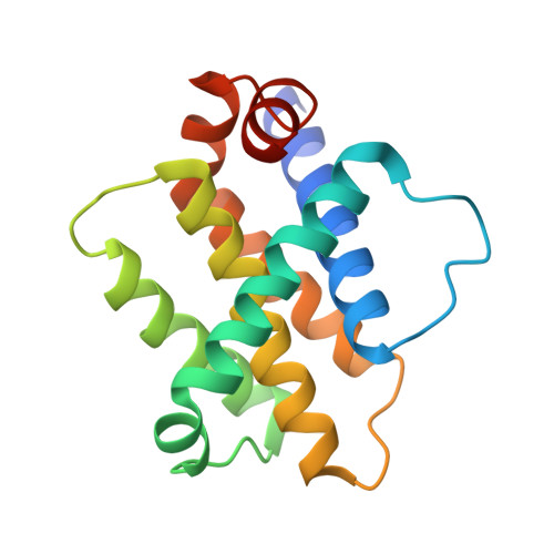
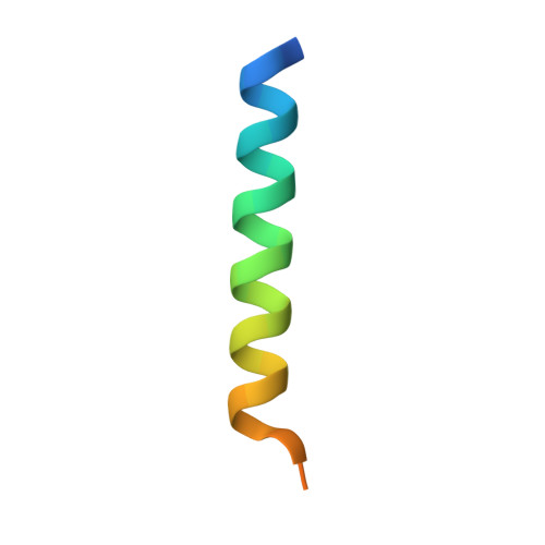
Structural Plasticity Underpins Promiscuous Binding of the Prosurvival Protein A1.

Smits, C.Czabotar, P.E.Hinds, M.G.Day, C.L.

(2008) Structure 16: 818

  • DOI: https://doi.org/10.1016/j.str.2008.02.009
  • Primary Citation Related Structures: 
    2VOF, 2VOG, 2VOH, 2VOI

  • PubMed Abstract: 

    Apoptotic pathways are regulated by protein-protein interactions. Interaction of the BH3 domains of proapoptotic Bcl-2 family proteins with the hydrophobic groove of prosurvival proteins is critical. Whereas some BH3 domains bind in a promiscuous manner, others exhibit considerable selectivity and the sequence characteristics that distinguish these activities are unclear. In this study, crystal structures of complexes between the prosurvival protein A1 and the BH3 domains from Puma, Bmf, Bak, and Bid have been solved. The structure of A1 is similar to that of other prosurvival proteins, although features, such as an acidic patch in the binding groove, may allow specific therapeutic modulation of apoptosis. Significant conformational plasticity was observed in the intermolecular interactions and these differences explain some of the variation in affinity. This study, in combination with published data, suggests that interactions between conserved residues demarcate optimal binding.


  • Organizational Affiliation
    • Biochemistry Department, University of Otago, Dunedin 9054, New Zealand.

Macromolecule Content 

  • Total Structure Weight: 21.9 kDa 
  • Atom Count: 1,407 
  • Modeled Residue Count: 174 
  • Deposited Residue Count: 191 
  • Unique protein chains: 2

Macromolecules

Find similar proteins by:|  3D Structure
Entity ID: 1
MoleculeChains  Sequence LengthOrganismDetailsImage
BCL-2-RELATED PROTEIN A1157Mus musculusMutation(s): 2 
UniProt
Find proteins for Q07440 (Mus musculus)
Explore Q07440 
Go to UniProtKB:  Q07440
Entity Groups
Sequence Clusters30% Identity50% Identity70% Identity90% Identity95% Identity100% Identity
UniProt GroupQ07440
Sequence Annotations
Expand
Reference Sequence
Find similar proteins by:|  3D Structure
Entity ID: 2
MoleculeChains  Sequence LengthOrganismDetailsImage
BH3-INTERACTING DOMAIN DEATH AGONIST P1334Mus musculusMutation(s): 0 
UniProt
Find proteins for P70444 (Mus musculus)
Explore P70444 
Go to UniProtKB:  P70444
Entity Groups
Sequence Clusters30% Identity50% Identity70% Identity90% Identity95% Identity100% Identity
UniProt GroupP70444
Sequence Annotations
Expand
Reference Sequence

Small Molecules

Ligands 1 Unique
IDChains Name / Formula / InChI Key2D Diagram3D Interactions
CL

Query on CL


Download:Ideal Coordinates CCD File
C [auth A]CHLORIDE ION
Cl
VEXZGXHMUGYJMC-UHFFFAOYSA-M

Experimental Data & Validation

Experimental Data

  • Method: X-RAY DIFFRACTION
  • Resolution: 2.10 Å
  • R-Value Free:  0.242 (Depositor), 0.249 (DCC) 
  • R-Value Work:  0.207 (Depositor), 0.210 (DCC) 
  • R-Value Observed: 0.209 (Depositor) 
Space Group: P 21 21 2
Unit Cell:
Length ( Å )Angle ( ˚ )
a = 61.737α = 90
b = 80.808β = 90
c = 32.184γ = 90
Software Package:
Software NamePurpose
REFMACrefinement
MOSFLMdata reduction
SCALAdata scaling
PHASERphasing

Structure Validation

View Full Validation Report


Entry History 

Deposition Data

Revision History  (Full details and data files)

  • Version 1.0: 2008-03-04
    Type: Initial release
  • Version 1.1: 2011-07-13
    Changes: Advisory, Refinement description, Version format compliance
  • Version 1.2: 2023-12-13
    Changes: Data collection, Database references, Other, Refinement description