2VO2 | pdb_00002vo2

Crystal structure of soybean ascorbate peroxidase mutant W41A subjected to low dose X-rays


Experimental Data Snapshot

  • Method: X-RAY DIFFRACTION
  • Resolution: 1.90 Å
  • R-Value Free: 
    0.236 (Depositor), 0.237 (DCC) 
  • R-Value Work: 
    0.174 (Depositor), 0.173 (DCC) 
  • R-Value Observed: 
    0.177 (Depositor) 

Starting Model: experimental
View more details

wwPDB Validation 3D Report Full Report

Validation slider image for 2VO2

Ligand Structure Quality Assessment 


This is version 1.5 of the entry. See complete history

Literature

Iron Oxidation State Modulates Active Site Structure in a Heme Peroxidase.

Badyal, S.K.Metcalfe, C.L.Basran, J.Efimov, I.Moody, P.C.E.Raven, E.L.

(2008) Biochemistry 47: 4403

  • DOI: https://doi.org/10.1021/bi702337n
  • Primary Citation Related Structures: 
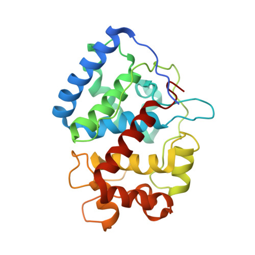
    2VNX, 2VNZ, 2VO2

  • PubMed Abstract: 

    We have previously shown [Badyal, S. K., et al. (2006) J. Biol. Chem. 281, 24512-24520] that the distal histidine (His42) in the W41A variant of ascorbate peroxidase binds to the heme iron in the ferric form of the protein but that binding of the substrate triggers a conformational change in which His42 dissociates from the heme. In this work, we show that this conformational rearrangement also occurs upon reduction of the heme iron. Thus, we present X-ray crystallographic data to show that reduction of the heme leads to dissociation of His42 from the iron in the ferrous form of W41A; spectroscopic and ligand binding data support this observation. Structural evidence indicates that heme reduction occurs through formation of a reduced, bis-histidine-ligated species that subsequently decays by dissociation of His42 from the heme. Collectively, the data provide clear evidence that conformational movement within the same heme active site can be controlled by both ligand binding and metal oxidation state. These observations are consistent with emerging data on other, more complex regulatory and sensing heme proteins, and the data are discussed in the context of our developing views in this area.


  • Organizational Affiliation
    • Department of Chemistry, Henry Wellcome Building, University of Leicester, UK.

Macromolecule Content 

  • Total Structure Weight: 28.98 kDa 
  • Atom Count: 2,265 
  • Modeled Residue Count: 248 
  • Deposited Residue Count: 261 
  • Unique protein chains: 1

Macromolecules

Find similar proteins by:|  3D Structure
Entity ID: 1
MoleculeChains  Sequence LengthOrganismDetailsImage
ASCORBATE PEROXIDASEA [auth X]261Glycine maxMutation(s): 1 
EC: 1.11.1.11
UniProt
Find proteins for Q43758 (Glycine max)
Explore Q43758 
Go to UniProtKB:  Q43758
Entity Groups
Sequence Clusters30% Identity50% Identity70% Identity90% Identity95% Identity100% Identity
UniProt GroupQ43758
Sequence Annotations
Expand
Reference Sequence

Experimental Data & Validation

Experimental Data

  • Method: X-RAY DIFFRACTION
  • Resolution: 1.90 Å
  • R-Value Free:  0.236 (Depositor), 0.237 (DCC) 
  • R-Value Work:  0.174 (Depositor), 0.173 (DCC) 
  • R-Value Observed: 0.177 (Depositor) 
Space Group: P 42 21 2
Unit Cell:
Length ( Å )Angle ( ˚ )
a = 82.057α = 90
b = 82.057β = 90
c = 75.631γ = 90
Software Package:
Software NamePurpose
REFMACrefinement
MOSFLMdata reduction
SCALAdata scaling
REFMACphasing

Structure Validation

View Full Validation Report


Ligand Structure Quality Assessment 


Entry History 

Deposition Data

Revision History  (Full details and data files)

  • Version 1.0: 2008-04-08
    Type: Initial release
  • Version 1.1: 2011-05-08
    Changes: Version format compliance
  • Version 1.2: 2011-07-13
    Changes: Version format compliance
  • Version 1.3: 2019-04-10
    Changes: Data collection, Other, Source and taxonomy
  • Version 1.4: 2024-05-08
    Changes: Data collection, Database references, Derived calculations, Other
  • Version 1.5: 2026-03-04
    Changes: Refinement description, Structure summary