2VLE | pdb_00002vle

The structure of daidzin, a naturally occurring anti alcohol- addiction agent, in complex with human mitochondrial aldehyde dehydrogenase


Experimental Data Snapshot

  • Method: X-RAY DIFFRACTION
  • Resolution: 2.40 Å
  • R-Value Free: 
    0.249 (Depositor), 0.240 (DCC) 
  • R-Value Work: 
    0.196 (Depositor), 0.190 (DCC) 

Starting Model: experimental
View more details

wwPDB Validation   3D Report Full Report


Ligand Structure Quality Assessment 

Created with Raphaël 2.3.0Worse 01 BetterLigand structure goodness of fit to experimental dataBest fitted DZNClick on this verticalbar to view details

This is version 1.3 of the entry. See complete history


Literature

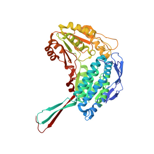
Structure of Daidzin, a Naturally Occurring Anti-Alcohol-Addiction Agent, in Complex with Human Mitochondrial Aldehyde Dehydrogenase.

Lowe, E.D.Gao, G.Y.Johnson, L.N.Keung, W.M.

(2008) J Med Chem 51: 4482

  • DOI: https://doi.org/10.1021/jm800488j
  • Primary Citation of Related Structures:  
    2VLE

  • PubMed Abstract: 

    The ALDH2*2 gene encoding the inactive variant form of mitochondrial aldehyde dehydrogenase (ALDH2) protects nearly all carriers of this gene from alcoholism. Inhibition of ALDH2 has hence become a possible strategy to treat alcoholism. The natural product 7-O-glucosyl-4'-hydroxyisoflavone (daidzin), isolated from the kudzu vine ( Peruraria lobata), is a specific inhibitor of ALDH2 and suppresses ethanol consumption. Daidzin is the active principle in a herbal remedy for "alcohol addiction" and provides a lead for the design of improved ALDH2. The structure of daidzin/ALDH2 in complex at 2.4 A resolution shows the isoflavone moiety of daidzin binding close to the aldehyde substrate-binding site in a hydrophobic cleft and the glucosyl function binding to a hydrophobic patch immediately outside the isoflavone-binding pocket. These observations provide an explanation for both the specificity and affinity of daidzin (IC50 =80 nM) and the affinity of analogues with different substituents at the glucosyl position.


  • Organizational Affiliation

    Laboratory of Molecular Biophysics, University of Oxford, Rex Richards Building, Oxford OX1 3QU, UK. edl@biop.ox.ac.uk


Macromolecules
Find similar proteins by:  (by identity cutoff)  |  3D Structure
Entity ID: 1
MoleculeChains Sequence LengthOrganismDetailsImage
ALDEHYDE DEHYDROGENASE, MITOCHONDRIAL
A, B, C, D, E
494Homo sapiensMutation(s): 0 
EC: 1.2.1.3
UniProt & NIH Common Fund Data Resources
Find proteins for P05091 (Homo sapiens)
Explore P05091 
Go to UniProtKB:  P05091
PHAROS:  P05091
GTEx:  ENSG00000111275 
Entity Groups  
Sequence Clusters30% Identity50% Identity70% Identity90% Identity95% Identity100% Identity
UniProt GroupP05091
Sequence Annotations
Expand
  • Reference Sequence
Small Molecules
Binding Affinity Annotations 
IDSourceBinding Affinity
DZN BindingDB:  2VLE IC50: min: 40, max: 3500 (nM) from 3 assay(s)
Experimental Data & Validation

Experimental Data

  • Method: X-RAY DIFFRACTION
  • Resolution: 2.40 Å
  • R-Value Free:  0.249 (Depositor), 0.240 (DCC) 
  • R-Value Work:  0.196 (Depositor), 0.190 (DCC) 
Space Group: P 21 21 21
Unit Cell:
Length ( Å )Angle ( ˚ )
a = 142.77α = 90
b = 150.99β = 90
c = 176.99γ = 90
Software Package:
Software NamePurpose
PHENIXrefinement
MOSFLMdata reduction
SCALAdata scaling
MOLREPphasing

Structure Validation

View Full Validation Report


Ligand Structure Quality Assessment 

Created with Raphaël 2.3.0Worse 01 BetterLigand structure goodness of fit to experimental dataBest fitted DZNClick on this verticalbar to view details

Entry History 

Deposition Data

Revision History  (Full details and data files)

  • Version 1.0: 2008-08-19
    Type: Initial release
  • Version 1.1: 2011-05-08
    Changes: Version format compliance
  • Version 1.2: 2011-07-13
    Changes: Version format compliance
  • Version 1.3: 2023-12-13
    Changes: Data collection, Database references, Other, Refinement description