2VLD | pdb_00002vld

crystal structure of a repair endonuclease from Pyrococcus abyssi


Experimental Data Snapshot

  • Method: X-RAY DIFFRACTION
  • Resolution: 2.60 Å
  • R-Value Free: 
    0.248 (Depositor), 0.239 (DCC) 
  • R-Value Work: 
    0.243 (Depositor), 0.232 (DCC) 
  • R-Value Observed: 
    0.243 (Depositor) 

wwPDB Validation 3D Report Full Report

Validation slider image for 2VLD

This is version 1.3 of the entry. See complete history

Literature

Structure and function of a novel endonuclease acting on branched DNA substrates.

Ren, B.Kuhn, J.Meslet-Cladiere, L.Briffotaux, J.Norais, C.Lavigne, R.Flament, D.Ladenstein, R.Myllykallio, H.

(2009) EMBO J 28: 2479-2489

  • DOI: https://doi.org/10.1038/emboj.2009.192
  • Primary Citation Related Structures: 
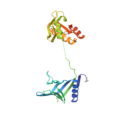
    2VLD

  • PubMed Abstract: 

    We show that Pyrococcus abyssi PAB2263 (dubbed NucS (nuclease for ss DNA) is a novel archaeal endonuclease that interacts with the replication clamp PCNA. Structural determination of P. abyssi NucS revealed a two-domain dumbbell-like structure that in overall does not resemble any known protein structure. Biochemical and structural studies indicate that NucS orthologues use a non-catalytic ssDNA-binding domain to regulate the cleavage activity at another site, thus resulting into the specific cleavage at double-stranded DNA (dsDNA)/ssDNA junctions on branched DNA substrates. Both 3' and 5' extremities of the ssDNA can be cleaved at the nuclease channel that is too narrow to accommodate duplex DNA. Altogether, our data suggest that NucS proteins constitute a new family of structure-specific DNA endonucleases that are widely distributed in archaea and in bacteria, including Mycobacterium tuberculosis.


  • Organizational Affiliation
    • Center for Structural Biochemistry, Karolinska Institutet, NOVUM, Huddinge, Sweden.

Macromolecule Content 

  • Total Structure Weight: 58.3 kDa 
  • Atom Count: 3,791 
  • Modeled Residue Count: 454 
  • Deposited Residue Count: 502 
  • Unique protein chains: 1

Macromolecules

Find similar proteins by:|  3D Structure
Entity ID: 1
MoleculeChains  Sequence LengthOrganismDetailsImage
Endonuclease NucS
A, B
251Pyrococcus abyssiMutation(s): 2 
Gene Names: nucSPYRAB01260PAB2263
EC: 3.1
UniProt
Find proteins for Q9V2E8 (Pyrococcus abyssi (strain GE5 / Orsay))
Explore Q9V2E8 
Go to UniProtKB:  Q9V2E8
Entity Groups
Sequence Clusters30% Identity50% Identity70% Identity90% Identity95% Identity100% Identity
UniProt GroupQ9V2E8
Sequence Annotations
Expand
Reference Sequence

Small Molecules

Modified Residues  1 Unique
IDChains TypeFormula2D DiagramParent
MSE
Query on MSE
A, B
L-PEPTIDE LINKINGC5 H11 N O2 SeMET

Experimental Data & Validation

Experimental Data

  • Method: X-RAY DIFFRACTION
  • Resolution: 2.60 Å
  • R-Value Free:  0.248 (Depositor), 0.239 (DCC) 
  • R-Value Work:  0.243 (Depositor), 0.232 (DCC) 
  • R-Value Observed: 0.243 (Depositor) 
Space Group: C 2 2 21
Unit Cell:
Length ( Å )Angle ( ˚ )
a = 78.966α = 90
b = 100.688β = 90
c = 157.686γ = 90
Software Package:
Software NamePurpose
REFMACrefinement
HKL-2000data reduction
HKL-2000data scaling
CNSphasing

Structure Validation

View Full Validation Report


Entry History 

Deposition Data

Revision History  (Full details and data files)

  • Version 1.0: 2009-05-19
    Type: Initial release
  • Version 1.1: 2011-07-13
    Changes: Advisory, Version format compliance
  • Version 1.2: 2018-09-26
    Changes: Data collection, Database references, Derived calculations, Source and taxonomy, Structure summary
  • Version 1.3: 2024-10-16
    Changes: Data collection, Database references, Derived calculations, Other, Refinement description, Structure summary