2VH5 | pdb_00002vh5

CRYSTAL STRUCTURE OF HRAS(G12V) - ANTI-RAS FV (disulfide free mutant) COMPLEX


Experimental Data Snapshot

  • Method: X-RAY DIFFRACTION
  • Resolution: 2.70 Å
  • R-Value Free: 
    0.291 (Depositor) 
  • R-Value Work: 
    0.211 (Depositor) 
  • R-Value Observed: 
    0.215 (Depositor) 

wwPDB Validation 3D Report Full Report

Validation slider image for 2VH5

This is version 1.4 of the entry. See complete history

Literature

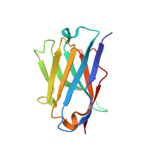
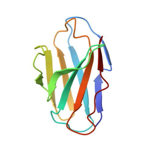
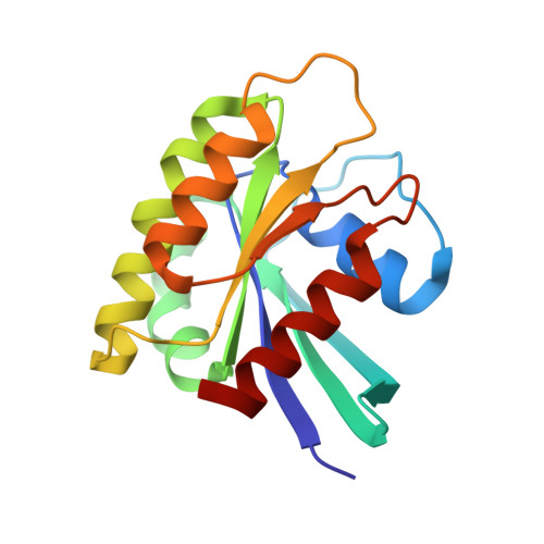
Functional Intracellular Antibody Fragments Do not Require Invariant Intra-Domain Disulfide Bonds.

Tanaka, T.Rabbitts, T.H.

(2008) J Mol Biology 376: 749

  • DOI: https://doi.org/10.1016/j.jmb.2007.11.085
  • Primary Citation Related Structures: 
    2VH5

  • PubMed Abstract: 

    Intracellular antibody fragments that interfere with molecular interactions inside cells are valuable in investigation of interactomes and in therapeutics, but their application demands that they function in the reducing cellular milieu. We show here a 2.7-A crystal structure of intracellular antibody folds based on scaffolds developed from intracellular antibody capture technology, and we reveal that there is no structural or functional difference with or without the intra-domain disulfide bond of the variable domain of heavy chain or the variable domain of light chain. The data indicate that, in the reducing in vivo environment, the absence of the intra-domain disulfide bond is not an impediment to correction of antibody folding or to interaction with antigen. Thus, the structural constraints for in-cell function are intrinsic to variable single-domain framework sequences, providing a generic scaffold for isolation of functional intracellular antibody single domains.


  • Organizational Affiliation
    • MRC Laboratory of Molecular Biology, Hills Road, Cambridge CB2 0QH, UK.

Macromolecule Content 

  • Total Structure Weight: 43.5 kDa 
  • Atom Count: 3,081 
  • Modeled Residue Count: 384 
  • Deposited Residue Count: 384 
  • Unique protein chains: 3

Macromolecules

Find similar proteins by:|  3D Structure
Entity ID: 1
MoleculeChains  Sequence LengthOrganismDetailsImage
ANTI-RAS FV HEAVY CHAINA [auth H]114Homo sapiensMutation(s): 0 
Find similar proteins by:|  3D Structure
Entity ID: 2
MoleculeChains  Sequence LengthOrganismDetailsImage
ANTI-RAS FV LIGHT CHAINB [auth L]104Homo sapiensMutation(s): 0 
Find similar proteins by:|  3D Structure
Entity ID: 3
MoleculeChains  Sequence LengthOrganismDetailsImage
GTPASE HRASC [auth R]166Homo sapiensMutation(s): 1 
EC: 3.6.5.2
UniProt & NIH Common Fund Data Resources
Find proteins for P01112 (Homo sapiens)
Explore P01112 
Go to UniProtKB:  P01112
PHAROS:  P01112
GTEx:  ENSG00000174775 
Entity Groups
Sequence Clusters30% Identity50% Identity70% Identity90% Identity95% Identity100% Identity
UniProt GroupP01112
Sequence Annotations
Expand
Reference Sequence

Experimental Data & Validation

Experimental Data

  • Method: X-RAY DIFFRACTION
  • Resolution: 2.70 Å
  • R-Value Free:  0.291 (Depositor) 
  • R-Value Work:  0.211 (Depositor) 
  • R-Value Observed: 0.215 (Depositor) 
Space Group: P 21 21 2
Unit Cell:
Length ( Å )Angle ( ˚ )
a = 75.863α = 90
b = 85.403β = 90
c = 63.084γ = 90
Software Package:
Software NamePurpose
REFMACrefinement
MOSFLMdata reduction
SCALAdata scaling

Structure Validation

View Full Validation Report


Entry History 

Deposition Data

Revision History  (Full details and data files)

  • Version 1.0: 2008-01-22
    Type: Initial release
  • Version 1.1: 2011-05-08
    Changes: Version format compliance
  • Version 1.2: 2011-07-13
    Changes: Version format compliance
  • Version 1.3: 2018-01-24
    Changes: Source and taxonomy
  • Version 1.4: 2024-05-08
    Changes: Data collection, Database references, Derived calculations