2VG6 | pdb_00002vg6

Crystal structures of HIV-1 reverse transcriptase complexes with thiocarbamate non-nucleoside inhibitors


Experimental Data Snapshot

  • Method: X-RAY DIFFRACTION
  • Resolution: 3.01 Å
  • R-Value Free: 
    0.335 (Depositor), 0.333 (DCC) 
  • R-Value Work: 
    0.244 (Depositor), 0.237 (DCC) 
  • R-Value Observed: 
    0.248 (Depositor) 

wwPDB Validation 3D Report Full Report

Validation slider image for 2VG6

Ligand Structure Quality Assessment 


This is version 1.4 of the entry. See complete history

Literature

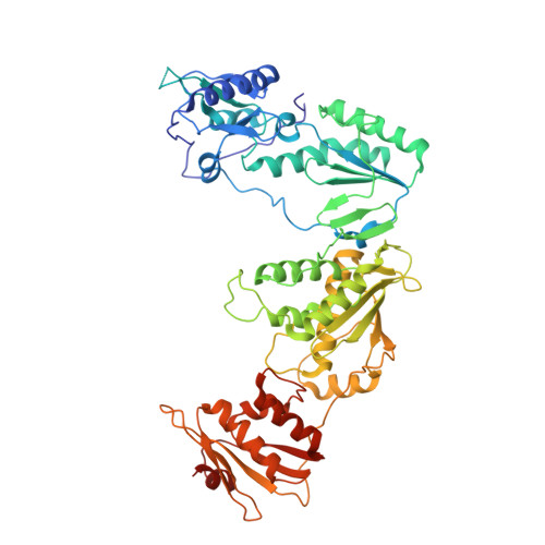
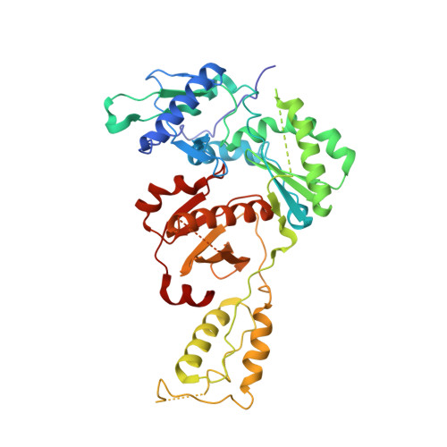
Crystal Structures of HIV-1 Reverse Transcriptase Complexes with Thiocarbamate Non-Nucleoside Inhibitors.

Spallarossa, A.Cesarini, S.Ranise, A.Ponassi, M.Unge, T.Bolognesi, M.

(2008) Biochem Biophys Res Commun 365: 764

  • DOI: https://doi.org/10.1016/j.bbrc.2007.11.036
  • Primary Citation Related Structures: 
    2VG5, 2VG6, 2VG7

  • PubMed Abstract: 

    O-Phthalimidoethyl-N-arylthiocarbamates (TCs) have been recently identified as a new class of potent HIV-1 reverse transcriptase (RT) non-nucleoside inhibitors (NNRTIs), by means of computer-aided drug design techniques [Ranise A. Spallarossa, S. Cesarini, F. Bondavalli, S. Schenone, O. Bruno, G. Menozzi, P. Fossa, L. Mosti, M. La Colla, et al., Structure-based design, parallel synthesis, structure-activity relationship, and molecular modeling studies of thiocarbamates, new potent non-nucleoside HIV-1 reverse transcriptase inhibitor isosteres of phenethylthiazolylthiourea derivatives, J. Med. Chem. 48 (2005) 3858-3873]. To elucidate the atomic details of RT/TC interaction and validate an earlier TC docking model, the structures of three RT/TC complexes were determined at 2.8-3.0A resolution by X-ray crystallography. The conformations adopted by the enzyme-bound TCs were analyzed and compared with those of bioisosterically related NNRTIs.


  • Organizational Affiliation
    • Dipartimento di Scienze Farmaceutiche, Universita' di Genova, viale Benedetto XV, 3, I-16132 Genova, Italy.

Macromolecule Content 

  • Total Structure Weight: 114.62 kDa 
  • Atom Count: 7,856 
  • Modeled Residue Count: 950 
  • Deposited Residue Count: 985 
  • Unique protein chains: 2

Macromolecules

Find similar proteins by:|  3D Structure
Entity ID: 1
MoleculeChains  Sequence LengthOrganismDetailsImage
REVERSE TRANSCRIPTASE/RIBONUCLEASE H557Human immunodeficiency virus 1Mutation(s): 0 
EC: 2.7.7.49 (PDB Primary Data), 2.7.7.7 (PDB Primary Data), 3.1.26.4 (PDB Primary Data)
UniProt
Find proteins for P03366 (Human immunodeficiency virus type 1 group M subtype B (isolate BH10))
Explore P03366 
Go to UniProtKB:  P03366
Entity Groups
Sequence Clusters30% Identity50% Identity70% Identity90% Identity95% Identity100% Identity
UniProt GroupP03366
Sequence Annotations
Expand
Reference Sequence
Find similar proteins by:|  3D Structure
Entity ID: 2
MoleculeChains  Sequence LengthOrganismDetailsImage
P51 RT428Human immunodeficiency virus 1Mutation(s): 0 
UniProt
Find proteins for P03366 (Human immunodeficiency virus type 1 group M subtype B (isolate BH10))
Explore P03366 
Go to UniProtKB:  P03366
Entity Groups
Sequence Clusters30% Identity50% Identity70% Identity90% Identity95% Identity100% Identity
UniProt GroupP03366
Sequence Annotations
Expand
Reference Sequence

Small Molecules

Ligands 1 Unique
IDChains Name / Formula / InChI Key2D Diagram3D Interactions
NNB

Query on NNB


Download:Ideal Coordinates CCD File
C [auth A]O-[2-(1,3-dioxo-1,3-dihydro-2H-isoindol-2-yl)ethyl] (4-bromophenyl)thiocarbamate
C17 H13 Br N2 O3 S
BDVAWAILQCYMQE-UHFFFAOYSA-N

Experimental Data & Validation

Experimental Data

  • Method: X-RAY DIFFRACTION
  • Resolution: 3.01 Å
  • R-Value Free:  0.335 (Depositor), 0.333 (DCC) 
  • R-Value Work:  0.244 (Depositor), 0.237 (DCC) 
  • R-Value Observed: 0.248 (Depositor) 
Space Group: C 2 2 21
Unit Cell:
Length ( Å )Angle ( ˚ )
a = 119.591α = 90
b = 157.183β = 90
c = 154.921γ = 90
Software Package:
Software NamePurpose
REFMACrefinement
AMoREphasing

Structure Validation

View Full Validation Report


Ligand Structure Quality Assessment 


Entry History 

Deposition Data

Revision History  (Full details and data files)

  • Version 1.0: 2007-12-11
    Type: Initial release
  • Version 1.1: 2011-05-08
    Changes: Version format compliance
  • Version 1.2: 2011-07-13
    Changes: Version format compliance
  • Version 1.3: 2018-01-17
    Changes: Data collection
  • Version 1.4: 2024-05-08
    Changes: Data collection, Database references, Other