2VE4 | pdb_00002ve4

Substrate free cyanobacterial CYP120A1


Experimental Data Snapshot

  • Method: X-RAY DIFFRACTION
  • Resolution: 2.40 Å
  • R-Value Free: 
    0.304 (Depositor), 0.303 (DCC) 
  • R-Value Work: 
    0.230 (Depositor), 0.230 (DCC) 
  • R-Value Observed: 
    0.234 (Depositor) 

wwPDB Validation 3D Report Full Report

Validation slider image for 2VE4

Ligand Structure Quality Assessment 


This is version 1.3 of the entry. See complete history

Literature

Crystal Structures of Substrate-Free and Retinoic Acid-Bound Cyanobacterial Cytochrome P450 Cyp120A1.

Kuhnel, K.Ke, N.Cryle, M.J.Sligar, S.G.Schuler, M.A.Schlichting, I.

(2008) Biochemistry 47: 6552

  • DOI: https://doi.org/10.1021/bi800328s
  • Primary Citation Related Structures: 
    2VE3, 2VE4

  • PubMed Abstract: 

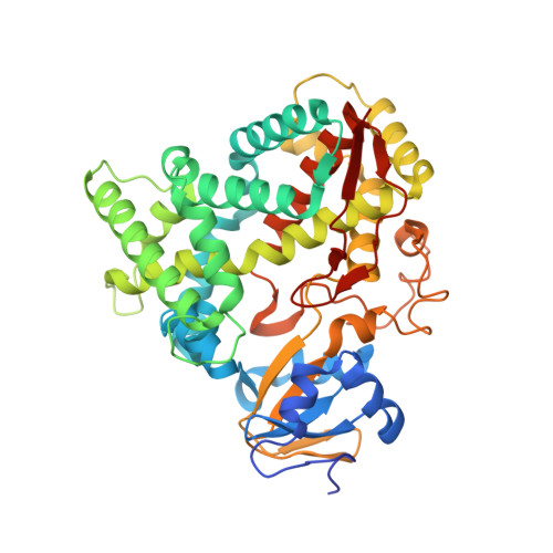
    The crystal structures of substrate-free and all-trans-retinoic acid-bound CYP120A1 from Synechocystis sp. PCC 6803 were determined at 2.4 and 2.1 A resolution, respectively, representing the first structural characterization of a cyanobacterial P450. Features of CYP120A1 not observed in other P450 structures include an aromatic ladder flanking the channel leading to the active site and a triple-glycine motif within SRS5. Using spectroscopic methods, CYP120A1 is shown to bind 13-cis-retinoic acid, 9-cis-retinoic acid, and retinal with high affinity and dissociation constants of less than 1 microM. Metabolism of retinoic acid by CYP120A1 suggests that CYP120A1 hydroxylates a variety of retinoid derivatives in vivo. On the basis of the retinoic acid-bound CYP120A1 crystal structure, we propose that either carbon 2 or the methyl groups (C16 or C17) of the beta-ionone ring are modified by CYP120A1.


  • Organizational Affiliation
    • Department of Biomolecular Mechanisms, Max Planck Institute for Medical Research, Jahnstrasse 29, 69120 Heidelberg, Germany.

Macromolecule Content 

  • Total Structure Weight: 102.5 kDa 
  • Atom Count: 7,144 
  • Modeled Residue Count: 863 
  • Deposited Residue Count: 888 
  • Unique protein chains: 1

Macromolecules

Find similar proteins by:|  3D Structure
Entity ID: 1
MoleculeChains  Sequence LengthOrganismDetailsImage
PUTATIVE CYTOCHROME P450 120
A, B
444Synechocystis sp. PCC 6803Mutation(s): 0 
EC: 1.14
UniProt
Find proteins for Q59990 (Synechocystis sp. (strain ATCC 27184 / PCC 6803 / Kazusa))
Explore Q59990 
Go to UniProtKB:  Q59990
Entity Groups
Sequence Clusters30% Identity50% Identity70% Identity90% Identity95% Identity100% Identity
UniProt GroupQ59990
Sequence Annotations
Expand
Reference Sequence

Experimental Data & Validation

Experimental Data

  • Method: X-RAY DIFFRACTION
  • Resolution: 2.40 Å
  • R-Value Free:  0.304 (Depositor), 0.303 (DCC) 
  • R-Value Work:  0.230 (Depositor), 0.230 (DCC) 
  • R-Value Observed: 0.234 (Depositor) 
Space Group: C 1 2 1
Unit Cell:
Length ( Å )Angle ( ˚ )
a = 153.64α = 90
b = 122.17β = 115
c = 64.92γ = 90
Software Package:
Software NamePurpose
REFMACrefinement
XDSdata reduction
XSCALEdata scaling

Structure Validation

View Full Validation Report


Ligand Structure Quality Assessment 


Entry History 

Deposition Data

Revision History  (Full details and data files)

  • Version 1.0: 2008-04-29
    Type: Initial release
  • Version 1.1: 2011-05-08
    Changes: Version format compliance
  • Version 1.2: 2011-07-13
    Changes: Version format compliance
  • Version 1.3: 2024-05-08
    Changes: Data collection, Database references, Derived calculations, Other