2V89 | pdb_00002v89

Crystal structure of RAG2-PHD finger in complex with H3K4me3 peptide at 1.1A resolution


Experimental Data Snapshot

  • Method: X-RAY DIFFRACTION
  • Resolution: 1.10 Å
  • R-Value Free: 
    0.155 (Depositor), 0.171 (DCC) 
  • R-Value Work: 
    0.157 (DCC) 
  • R-Value Observed: 
    0.123 (Depositor) 

Starting Model: experimental
View more details

wwPDB Validation 3D Report Full Report

Validation slider image for 2V89

This is version 2.0 of the entry. See complete history

Literature

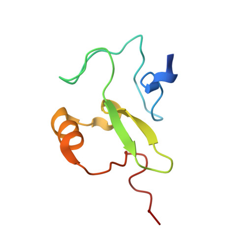
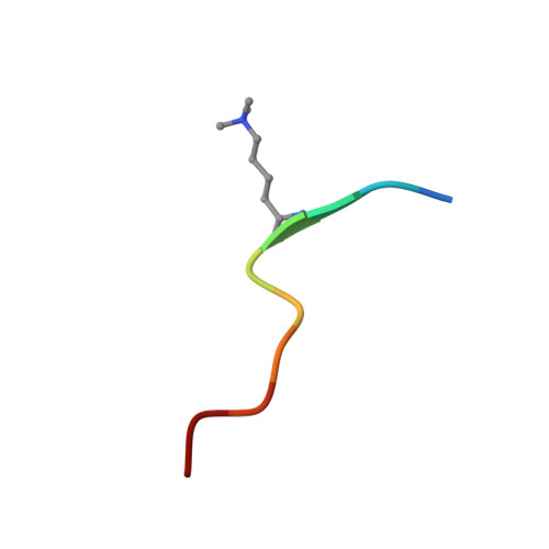
Rag2 Phd Finger Couples Histone H3 Lysine 4 Trimethylation with V(D)J Recombination.

Matthews, A.G.W.Kuo, A.J.Ramon-Maiques, S.Han, S.Champagne, K.S.Ivanov, D.Gallardo, M.Carney, D.Cheung, P.Ciccone, D.N.Walter, K.L.Utz, P.J.Shi, Y.Kutateladze, T.G.Yang, W.Gozani, O.Oettinger, M.A.

(2007) Nature 450: 1106

  • DOI: https://doi.org/10.1038/nature06431
  • Primary Citation Related Structures: 
    2V89

  • PubMed Abstract: 

    Nuclear processes such as transcription, DNA replication and recombination are dynamically regulated by chromatin structure. Eukaryotic transcription is known to be regulated by chromatin-associated proteins containing conserved protein domains that specifically recognize distinct covalent post-translational modifications on histones. However, it has been unclear whether similar mechanisms are involved in mammalian DNA recombination. Here we show that RAG2--an essential component of the RAG1/2 V(D)J recombinase, which mediates antigen-receptor gene assembly--contains a plant homeodomain (PHD) finger that specifically recognizes histone H3 trimethylated at lysine 4 (H3K4me3). The high-resolution crystal structure of the mouse RAG2 PHD finger bound to H3K4me3 reveals the molecular basis of H3K4me3-recognition by RAG2. Mutations that abrogate RAG2's recognition of H3K4me3 severely impair V(D)J recombination in vivo. Reducing the level of H3K4me3 similarly leads to a decrease in V(D)J recombination in vivo. Notably, a conserved tryptophan residue (W453) that constitutes a key structural component of the K4me3-binding surface and is essential for RAG2's recognition of H3K4me3 is mutated in patients with immunodeficiency syndromes. Together, our results identify a new function for histone methylation in mammalian DNA recombination. Furthermore, our results provide the first evidence indicating that disrupting the read-out of histone modifications can cause an inherited human disease.


  • Organizational Affiliation
    • Department of Molecular Biology, Massachusetts General Hospital, Harvard Medical School, Boston, Massachusetts 02114, USA.

Macromolecule Content 

  • Total Structure Weight: 21.16 kDa 
  • Atom Count: 1,728 
  • Modeled Residue Count: 172 
  • Deposited Residue Count: 184 
  • Unique protein chains: 2

Macromolecules

Find similar proteins by:|  3D Structure
Entity ID: 1
MoleculeChains  Sequence LengthOrganismDetailsImage
VDJ RECOMBINATION-ACTIVATING PROTEIN 2
A, B
82Mus musculusMutation(s): 0 
UniProt
Find proteins for P21784 (Mus musculus)
Explore P21784 
Go to UniProtKB:  P21784
Entity Groups
Sequence Clusters30% Identity50% Identity70% Identity90% Identity95% Identity100% Identity
UniProt GroupP21784
Sequence Annotations
Expand
Reference Sequence
Find similar proteins by:  Sequence   |   3D Structure  
Entity ID: 2
MoleculeChains  Sequence LengthOrganismDetailsImage
HISTONE H3C [auth D],
D [auth E]
10Homo sapiensMutation(s): 0 
UniProt & NIH Common Fund Data Resources
Find proteins for P68431 (Homo sapiens)
Explore P68431 
Go to UniProtKB:  P68431
Entity Groups
Sequence Clusters30% Identity50% Identity70% Identity90% Identity95% Identity100% Identity
UniProt GroupP68431
Sequence Annotations
Expand
Reference Sequence

Experimental Data & Validation

Experimental Data

  • Method: X-RAY DIFFRACTION
  • Resolution: 1.10 Å
  • R-Value Free:  0.155 (Depositor), 0.171 (DCC) 
  • R-Value Work:  0.157 (DCC) 
  • R-Value Observed: 0.123 (Depositor) 
Space Group: C 1 2 1
Unit Cell:
Length ( Å )Angle ( ˚ )
a = 75.978α = 90
b = 46.807β = 106.78
c = 56.808γ = 90
Software Package:
Software NamePurpose
SHELXL-97refinement
HKL-2000data reduction
HKL-2000data scaling
PHASERphasing

Structure Validation

View Full Validation Report


Entry History 

Deposition Data

Revision History  (Full details and data files)

  • Version 1.0: 2007-12-11
    Type: Initial release
  • Version 1.1: 2013-03-13
    Changes: Database references, Derived calculations, Non-polymer description, Other, Structure summary, Version format compliance
  • Version 1.2: 2016-12-28
    Changes: Database references, Other, Source and taxonomy, Structure summary
  • Version 1.3: 2019-03-06
    Changes: Data collection, Derived calculations, Experimental preparation, Other
  • Version 1.4: 2019-05-08
    Changes: Data collection, Experimental preparation
  • Version 2.0: 2023-12-13
    Changes: Atomic model, Data collection, Database references, Derived calculations, Other, Refinement description