2V79 | pdb_00002v79

Crystal Structure of the N-terminal domain of DnaD from Bacillus Subtilis


Experimental Data Snapshot

  • Method: X-RAY DIFFRACTION
  • Resolution: 2.00 Å
  • R-Value Free: 
    0.210 (Depositor), 0.218 (DCC) 
  • R-Value Work: 
    0.181 (Depositor), 0.196 (DCC) 
  • R-Value Observed: 
    0.182 (Depositor) 

wwPDB Validation 3D Report Full Report

Validation slider image for 2V79

This is version 1.3 of the entry. See complete history

Literature

Structure of the N-Terminal Oligomerization Domain of Dnad Reveals a Unique Tetramerization Motif and Provides Insights Into Scaffold Formation.

Schneider, S.Zhang, W.Soultanas, P.Paoli, M.

(2008) J Mol Biology 376: 1237

  • DOI: https://doi.org/10.1016/j.jmb.2007.12.045
  • Primary Citation Related Structures: 
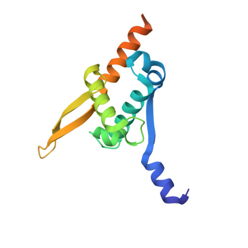
    2V79

  • PubMed Abstract: 

    DnaD is a primosomal protein that remodels supercoiled plasmids. It binds to supercoiled forms and converts them to open forms without nicking. During this remodeling process, all the writhe is converted to twist and the plasmids are held around the periphery of large scaffolds made up of DnaD molecules. This DNA-remodeling function is the sum of a scaffold-forming activity on the N-terminal domain and a DNA-dependent oligomerization activity on the C-terminal domain. We have determined the crystal structure of the scaffold-forming N-terminal domain, which reveals a winged-helix architecture, with additional structural elements extending from both N- and C-termini. Four monomers form dimers that join into a tetramer. The N-terminal extension mediates dimerization and tetramerization, with extensive interactions and distinct interfaces. The wings and helices of the winged-helix domains remain exposed on the surface of the tetramer. Structure-guided mutagenesis and atomic force microscopy imaging indicate that these elements, together with the C-terminal extension, are involved in scaffold formation. Based upon our data, we propose a model for the DnaD-mediated scaffold formation.


  • Organizational Affiliation
    • Centre for Biomolecular Sciences, University of Nottingham, University Park, Nottingham NG7 2RD, UK.

Macromolecule Content 

  • Total Structure Weight: 32.62 kDa 
  • Atom Count: 2,189 
  • Modeled Residue Count: 232 
  • Deposited Residue Count: 270 
  • Unique protein chains: 1

Macromolecules

Find similar proteins by:|  3D Structure
Entity ID: 1
MoleculeChains  Sequence LengthOrganismDetailsImage
DNA REPLICATION PROTEIN DNAD
A, B
135Bacillus subtilisMutation(s): 0 
UniProt
Find proteins for P39787 (Bacillus subtilis (strain 168))
Explore P39787 
Go to UniProtKB:  P39787
Entity Groups
Sequence Clusters30% Identity50% Identity70% Identity90% Identity95% Identity100% Identity
UniProt GroupP39787
Sequence Annotations
Expand
Reference Sequence

Small Molecules

Ligands 2 Unique
IDChains Name / Formula / InChI Key2D Diagram3D Interactions
CL

Query on CL


Download:Ideal Coordinates CCD File
E [auth A]
F [auth A]
G [auth A]
H [auth A]
J [auth B]
E [auth A],
F [auth A],
G [auth A],
H [auth A],
J [auth B],
K [auth B],
L [auth B],
M [auth B],
N [auth B],
O [auth B],
P [auth B]
CHLORIDE ION
Cl
VEXZGXHMUGYJMC-UHFFFAOYSA-M
NA

Query on NA


Download:Ideal Coordinates CCD File
C [auth A],
D [auth A],
I [auth B]
SODIUM ION
Na
FKNQFGJONOIPTF-UHFFFAOYSA-N

Experimental Data & Validation

Experimental Data

  • Method: X-RAY DIFFRACTION
  • Resolution: 2.00 Å
  • R-Value Free:  0.210 (Depositor), 0.218 (DCC) 
  • R-Value Work:  0.181 (Depositor), 0.196 (DCC) 
  • R-Value Observed: 0.182 (Depositor) 
Space Group: P 31 2 1
Unit Cell:
Length ( Å )Angle ( ˚ )
a = 78.666α = 90
b = 78.666β = 90
c = 124.558γ = 120
Software Package:
Software NamePurpose
REFMACrefinement
MOSFLMdata reduction
SCALAdata scaling
SHARPphasing

Structure Validation

View Full Validation Report


Entry History 

Deposition Data

Revision History  (Full details and data files)

  • Version 1.0: 2008-01-15
    Type: Initial release
  • Version 1.1: 2011-07-13
    Changes: Refinement description, Version format compliance
  • Version 1.2: 2017-06-28
    Changes: Data collection
  • Version 1.3: 2024-05-08
    Changes: Advisory, Data collection, Database references, Derived calculations, Other