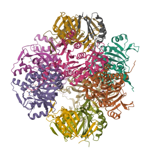
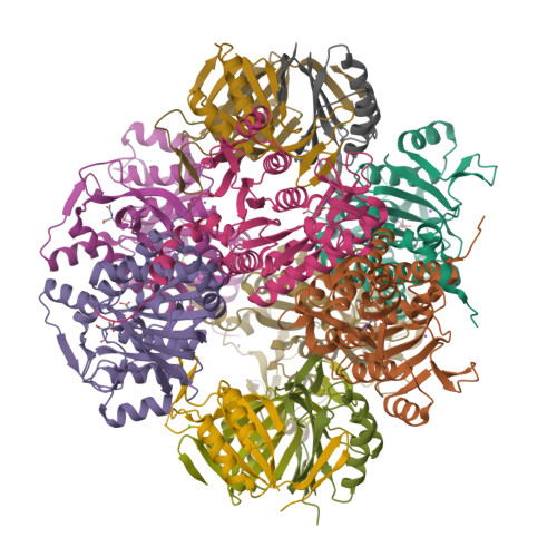
2V5H

Controlling the storage of nitrogen as arginine: the complex of PII and acetylglutamate kinase from Synechococcus elongatus PCC 7942


Experimental Data Snapshot

  • Method: X-RAY DIFFRACTION
  • Resolution: 2.75 Å
  • R-Value Free: 0.236 
  • R-Value Work: 0.200 
  • R-Value Observed: 0.202 

Starting Model: experimental
View more details

wwPDB Validation   3D Report Full Report


Ligand Structure Quality Assessment 


This is version 1.3 of the entry. See complete history


Literature

The Crystal Structure of the Complex of Pii and Acetylglutamate Kinase Reveals How Pii Controls the Storage of Nitrogen as Arginine.

Llacer, J.L.Contreras, A.Forchhammer, K.Marco-Marin, C.Gil-Ortiz, F.Maldonado, R.Fita, I.Rubio, V.

(2007) Proc Natl Acad Sci U S A 104: 17644

  • DOI: https://doi.org/10.1073/pnas.0705987104
  • Primary Citation of Related Structures:  
    2JJ4, 2V5H

  • PubMed Abstract: 

    Photosynthetic organisms can store nitrogen by synthesizing arginine, and, therefore, feedback inhibition of arginine synthesis must be relieved in these organisms when nitrogen is abundant. This relief is accomplished by the binding of the PII signal transduction protein to acetylglutamate kinase (NAGK), the controlling enzyme of arginine synthesis. Here, we describe the crystal structure of the complex between NAGK and PII of Synechococcus elongatus, at 2.75-A resolution. We prove the physiological relevance of the observed interactions by site-directed mutagenesis and functional studies. The complex consists of two polar PII trimers sandwiching one ring-like hexameric NAGK (a trimer of dimers) with the threefold axes of these molecules aligned. The binding of PII favors a narrow ring conformation of the NAGK hexamer that is associated with arginine sites having low affinity for this inhibitor. Each PII subunit contacts one NAGK subunit only. The contacts map in the inner circumference of the NAGK ring and involve two surfaces of the PII subunit. One surface is on the PII body and interacts with the C-domain of the NAGK subunit, helping widen the arginine site found on the other side of this domain. The other surface is at the distal region of a protruding large loop (T-loop) that presents a novel compact shape. This loop is inserted in the interdomain crevice of the NAGK subunit, contacting mainly the N-domain, and playing key roles in anchoring PII on NAGK, in activating NAGK, and in complex formation regulation by MgATP, ADP, 2-oxoglutarate, and by phosphorylation of serine-49.


  • Organizational Affiliation

    Instituto de Biomedicina de Valencia-Consejo Superior de Investigaciones Cientificas and Centro de Investigación Biomédica en Red de Enfermedades Raras, Jaime Roig 11, 46010 Valencia, Spain.


Macromolecules
Find similar proteins by:  (by identity cutoff)  |  3D Structure
Entity ID: 1
MoleculeChains Sequence LengthOrganismDetailsImage
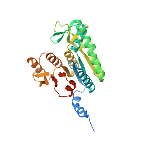
ACETYLGLUTAMATE KINASE
A, B, C, D, E
A, B, C, D, E, F
321Synechococcus elongatus PCC 7942 = FACHB-805Mutation(s): 0 
EC: 2.7.2.8
UniProt
Find proteins for Q6V1L5 (Synechococcus elongatus (strain ATCC 33912 / PCC 7942 / FACHB-805))
Explore Q6V1L5 
Go to UniProtKB:  Q6V1L5
Entity Groups  
Sequence Clusters30% Identity50% Identity70% Identity90% Identity95% Identity100% Identity
UniProt GroupQ6V1L5
Sequence Annotations
Expand
  • Reference Sequence
Find similar proteins by:  (by identity cutoff)  |  3D Structure
Entity ID: 2
MoleculeChains Sequence LengthOrganismDetailsImage
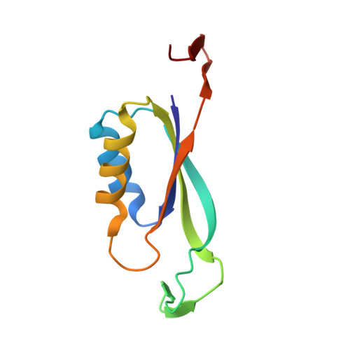
NITROGEN REGULATORY PROTEIN P-II
G, H, I, J, K
G, H, I, J, K, L
112Synechococcus elongatus PCC 7942 = FACHB-805Mutation(s): 0 
UniProt
Find proteins for P0A3F4 (Synechococcus elongatus (strain ATCC 33912 / PCC 7942 / FACHB-805))
Explore P0A3F4 
Go to UniProtKB:  P0A3F4
Entity Groups  
Sequence Clusters30% Identity50% Identity70% Identity90% Identity95% Identity100% Identity
UniProt GroupP0A3F4
Sequence Annotations
Expand
  • Reference Sequence
Small Molecules
Ligands 4 Unique
IDChains Name / Formula / InChI Key2D Diagram3D Interactions
NLG
Query on NLG

Download Ideal Coordinates CCD File 
M [auth A]
O [auth B]
Q [auth C]
S [auth D]
U [auth E]
M [auth A],
O [auth B],
Q [auth C],
S [auth D],
U [auth E],
X [auth F]
N-ACETYL-L-GLUTAMATE
C7 H11 N O5
RFMMMVDNIPUKGG-YFKPBYRVSA-N
GOL
Query on GOL

Download Ideal Coordinates CCD File 
V [auth E]GLYCEROL
C3 H8 O3
PEDCQBHIVMGVHV-UHFFFAOYSA-N
CL
Query on CL

Download Ideal Coordinates CCD File 
AA [auth H]
BA [auth H]
CA [auth J]
DA [auth J]
EA [auth K]
AA [auth H],
BA [auth H],
CA [auth J],
DA [auth J],
EA [auth K],
Z [auth G]
CHLORIDE ION
Cl
VEXZGXHMUGYJMC-UHFFFAOYSA-M
NA
Query on NA

Download Ideal Coordinates CCD File 
N [auth A]
P [auth B]
R [auth C]
T [auth D]
W [auth E]
N [auth A],
P [auth B],
R [auth C],
T [auth D],
W [auth E],
Y [auth F]
SODIUM ION
Na
FKNQFGJONOIPTF-UHFFFAOYSA-N
Experimental Data & Validation

Experimental Data

  • Method: X-RAY DIFFRACTION
  • Resolution: 2.75 Å
  • R-Value Free: 0.236 
  • R-Value Work: 0.200 
  • R-Value Observed: 0.202 
  • Space Group: P 1 21 1
Unit Cell:
Length ( Å )Angle ( ˚ )
a = 90.379α = 90
b = 161.027β = 106.53
c = 91.562γ = 90
Software Package:
Software NamePurpose
REFMACrefinement
MOSFLMdata reduction
SCALAdata scaling
MOLREPphasing

Structure Validation

View Full Validation Report


Ligand Structure Quality Assessment 


Entry History 

Deposition Data

Revision History  (Full details and data files)

  • Version 1.0: 2007-10-16
    Type: Initial release
  • Version 1.1: 2011-07-13
    Changes: Advisory, Version format compliance
  • Version 1.2: 2014-12-10
    Changes: Data collection, Database references, Derived calculations, Non-polymer description
  • Version 1.3: 2023-12-13
    Changes: Data collection, Database references, Derived calculations, Other, Refinement description