2V23 | pdb_00002v23

Structure of cytochrome c peroxidase mutant N184R Y36A


Experimental Data Snapshot

  • Method: X-RAY DIFFRACTION
  • Resolution: 1.80 Å
  • R-Value Free: 
    0.192 (Depositor), 0.191 (DCC) 
  • R-Value Work: 
    0.159 (Depositor), 0.159 (DCC) 
  • R-Value Observed: 
    0.161 (Depositor) 

Starting Model: experimental
View more details

wwPDB Validation 3D Report Full Report

Validation slider image for 2V23

Ligand Structure Quality Assessment 


This is version 1.5 of the entry. See complete history

Literature

The Tuberculosis Prodrug Isoniazid Bound to Activating Peroxidases.

Metcalfe, C.L.Macdonald, I.K.Murphy, E.J.Brown, K.A.Raven, E.L.Moody, P.C.E.

(2008) J Biological Chem 283: 6193

  • DOI: https://doi.org/10.1074/jbc.M707412200
  • Primary Citation Related Structures: 
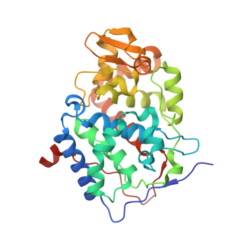
    2V23, 2V2E, 2VCF, 2VCN, 2VCS

  • PubMed Abstract: 

    Isoniazid (INH, isonicotinic acid hydrazine) is one of only two therapeutic agents effective in treating tuberculosis. This prodrug is activated by the heme enzyme catalase peroxidase (KatG) endogenous to Mycobacterium tuberculosis but the mechanism of activation is poorly understood, in part because the binding interaction has not been properly established. The class I peroxidases ascorbate peroxidase (APX) and cytochrome c peroxidase (CcP) have active site structures very similar to KatG and are also capable of activating isoniazid. We report here the first crystal structures of complexes of isoniazid bound to APX and CcP. These are the first structures of isoniazid bound to any activating enzymes. The structures show that isoniazid binds close to the delta-heme edge in both APX and CcP, although the precise binding orientation varies slightly in the two cases. A second binding site for INH is found in APX at the gamma-heme edge close to the established ascorbate binding site, indicating that the gamma-heme edge can also support the binding of aromatic substrates. We also show that in an active site mutant of soybean APX (W41A) INH can bind directly to the heme iron to become an inhibitor and in a different mode when the distal histidine is replaced by alanine (H42A). These structures provide the first unambiguous evidence for the location of the isoniazid binding site in the class I peroxidases and provide rationalization of isoniazid resistance in naturally occurring KatG mutant strains of M. tuberculosis.


  • Organizational Affiliation
    • Department of Chemistry, University of Leicester, Lancaster Road, Leicester, United Kingdom.

Macromolecule Content 

  • Total Structure Weight: 34.4 kDa 
  • Atom Count: 2,766 
  • Modeled Residue Count: 293 
  • Deposited Residue Count: 296 
  • Unique protein chains: 1

Macromolecules

Find similar proteins by:|  3D Structure
Entity ID: 1
MoleculeChains  Sequence LengthOrganismDetailsImage
CYTOCHROME C PEROXIDASE296Saccharomyces cerevisiaeMutation(s): 2 
EC: 1.11.1.5
UniProt
Find proteins for P00431 (Saccharomyces cerevisiae (strain ATCC 204508 / S288c))
Explore P00431 
Go to UniProtKB:  P00431
Entity Groups
Sequence Clusters30% Identity50% Identity70% Identity90% Identity95% Identity100% Identity
UniProt GroupP00431
Sequence Annotations
Expand
Reference Sequence

Small Molecules

Ligands 1 Unique
IDChains Name / Formula / InChI Key2D Diagram3D Interactions
HEM

Query on HEM


Download:Ideal Coordinates CCD File
B [auth A]PROTOPORPHYRIN IX CONTAINING FE
C34 H32 Fe N4 O4
KABFMIBPWCXCRK-RGGAHWMASA-L

Experimental Data & Validation

Experimental Data

  • Method: X-RAY DIFFRACTION
  • Resolution: 1.80 Å
  • R-Value Free:  0.192 (Depositor), 0.191 (DCC) 
  • R-Value Work:  0.159 (Depositor), 0.159 (DCC) 
  • R-Value Observed: 0.161 (Depositor) 
Space Group: P 21 21 21
Unit Cell:
Length ( Å )Angle ( ˚ )
a = 51.146α = 90
b = 75.377β = 90
c = 107.228γ = 90
Software Package:
Software NamePurpose
REFMACrefinement
MOSFLMdata reduction
SCALAdata scaling
REFMACphasing

Structure Validation

View Full Validation Report


Ligand Structure Quality Assessment 


Entry History 

Deposition Data

Revision History  (Full details and data files)

  • Version 1.0: 2007-12-04
    Type: Initial release
  • Version 1.1: 2011-05-08
    Changes: Version format compliance
  • Version 1.2: 2011-07-13
    Changes: Version format compliance
  • Version 1.3: 2017-07-05
    Changes: Data collection
  • Version 1.4: 2019-01-30
    Changes: Data collection, Experimental preparation
  • Version 1.5: 2023-12-13
    Changes: Data collection, Database references, Derived calculations, Other, Refinement description