2V17 | pdb_00002v17

Structure of the complex of antibody MN423 with a fragment of tau protein


Experimental Data Snapshot

  • Method: X-RAY DIFFRACTION
  • Resolution: 1.65 Å
  • R-Value Free: 
    0.218 (Depositor), 0.228 (DCC) 
  • R-Value Work: 
    0.156 (Depositor), 0.170 (DCC) 
  • R-Value Observed: 
    0.160 (Depositor) 

Starting Model: experimental
View more details

wwPDB Validation 3D Report Full Report

Validation slider image for 2V17

This is version 1.5 of the entry. See complete history

Literature

X-Ray Structure of the Phf Core C-Terminus: Insight Into the Folding of the Intrinsically Disordered Protein Tau in Alzheimer'S Disease.

Sevcik, J.Skrabana, R.Dvorsky, R.Csokova, N.Iqbal, K.Novak, M.

(2007) FEBS Lett 581: 5872

  • DOI: https://doi.org/10.1016/j.febslet.2007.11.067
  • Primary Citation Related Structures: 
    2V17

  • PubMed Abstract: 

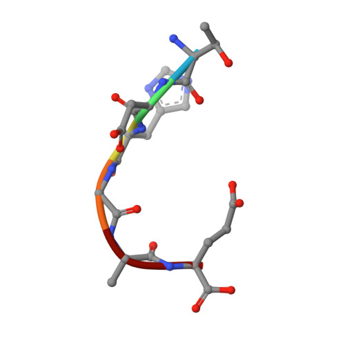
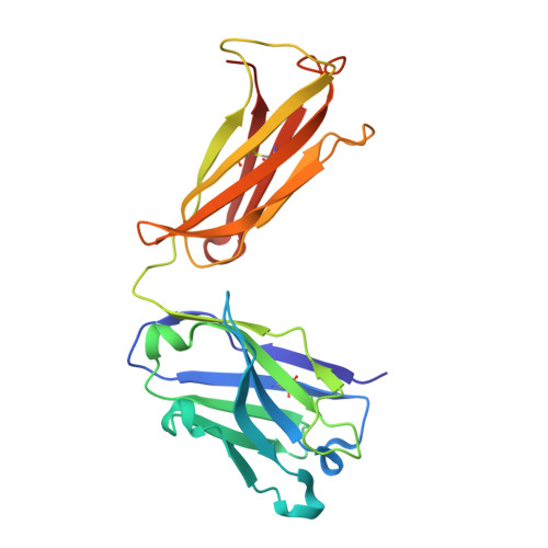
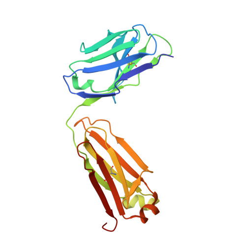
    The major constituent of Alzheimer's disease paired helical filaments (PHF) core is intrinsically disordered protein (IDP) tau. In spite of a considerable effort, insoluble character of PHF together with inherent physical properties of IDP tau have precluded so far reconstruction of PHF 3D structure by X-ray crystallography or NMR spectroscopy. Here we present first crystallographic study of PHF core C-terminus. Using monoclonal antibody MN423 specific to the tertiary structure of the PHF core, the in vivo PHF structure was imprinted into recombinant core PHF tau. Crystallization of the complex led to determination of the structure of the core PHF tau protein fragment 386TDHGAE391 at 1.65A resolution. Structural analysis suggests important role of the core PHF C-terminus for PHF assembly. It is reasonable to expect that this approach will help to reveal the structural principles underlying the tau protein assembly into PHF and possibly will facilitate rationale drug design for inhibition of Alzheimer neurofibrillary changes.


  • Organizational Affiliation
    • Institute of Neuroimmunology, Slovak Academy of Sciences, Dubravska cesta 9, 845 10 Bratislava, Slovakia.

Macromolecule Content 

  • Total Structure Weight: 47.91 kDa 
  • Atom Count: 4,069 
  • Modeled Residue Count: 442 
  • Deposited Residue Count: 442 
  • Unique protein chains: 3

Macromolecules

Find similar proteins by:  Sequence   |   3D Structure  
Entity ID: 1
MoleculeChains  Sequence LengthOrganismDetailsImage
PEPTIDE FRAGMENT6Homo sapiensMutation(s): 0 
Find similar proteins by:|  3D Structure
Entity ID: 2
MoleculeChains  Sequence LengthOrganismDetailsImage
MONOCLONAL ANTIBODY FAB FRAGMENT MN423B [auth H]222Mus musculusMutation(s): 0 
Find similar proteins by:|  3D Structure
Entity ID: 3
MoleculeChains  Sequence LengthOrganismDetailsImage
MONOCLONAL ANTIBODY FAB FRAGMENT MN423C [auth L]214Mus musculusMutation(s): 0 
UniProt
Find proteins for P01837 (Mus musculus)
Explore P01837 
Go to UniProtKB:  P01837
Entity Groups
Sequence Clusters30% Identity50% Identity70% Identity90% Identity95% Identity100% Identity
UniProt GroupP01837
Sequence Annotations
Expand
Reference Sequence

Experimental Data & Validation

Experimental Data

  • Method: X-RAY DIFFRACTION
  • Resolution: 1.65 Å
  • R-Value Free:  0.218 (Depositor), 0.228 (DCC) 
  • R-Value Work:  0.156 (Depositor), 0.170 (DCC) 
  • R-Value Observed: 0.160 (Depositor) 
Space Group: P 1 21 1
Unit Cell:
Length ( Å )Angle ( ˚ )
a = 71.541α = 90
b = 36.808β = 113.93
c = 85.543γ = 90
Software Package:
Software NamePurpose
REFMACrefinement
DENZOdata reduction
SCALEPACKdata scaling
MOLREPphasing

Structure Validation

View Full Validation Report


Entry History 

Deposition Data

Revision History  (Full details and data files)

  • Version 1.0: 2007-12-25
    Type: Initial release
  • Version 1.1: 2011-05-08
    Changes: Version format compliance
  • Version 1.2: 2011-07-13
    Changes: Version format compliance
  • Version 1.3: 2019-07-24
    Changes: Data collection
  • Version 1.4: 2023-12-13
    Changes: Data collection, Database references, Other, Refinement description
  • Version 1.5: 2024-10-23
    Changes: Structure summary