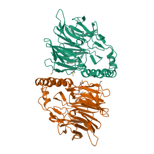
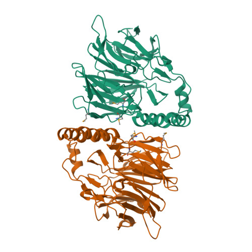
2UVK

Structure of YjhT


Experimental Data Snapshot

  • Method: X-RAY DIFFRACTION
  • Resolution: 1.50 Å
  • R-Value Free: 0.219 
  • R-Value Work: 0.180 
  • R-Value Observed: 0.182 

wwPDB Validation   3D Report Full Report


This is version 1.3 of the entry. See complete history


Literature

Sialic Acid Mutarotation is Catalyzed by the Escherichia Coli Beta-Propeller Protein Yjht.

Severi, E.Muller, A.Potts, J.R.Leech, A.Williamson, D.Wilson, K.S.Thomas, G.H.

(2008) J Biol Chem 283: 4841

  • DOI: https://doi.org/10.1074/jbc.M707822200
  • Primary Citation of Related Structures:  
    2UVK

  • PubMed Abstract: 

    The acquisition of host-derived sialic acid is an important virulence factor for some bacterial pathogens, but in vivo this sugar acid is sequestered in sialoconjugates as the alpha-anomer. In solution, however, sialic acid is present mainly as the beta-anomer, formed by a slow spontaneous mutarotation. We studied the Escherichia coli protein YjhT as a member of a family of uncharacterized proteins present in many sialic acid-utilizing pathogens. This protein is able to accelerate the equilibration of the alpha- and beta-anomers of the sialic acid N-acetylneuraminic acid, thus describing a novel sialic acid mutarotase activity. The structure of this periplasmic protein, solved to 1.5A resolution, reveals a dimeric 6-bladed unclosed beta-propeller, the first of a bacterial Kelch domain protein. Mutagenesis of conserved residues in YjhT demonstrated an important role for Glu-209 and Arg-215 in mutarotase activity. We also present data suggesting that the ability to utilize alpha-N-acetylneuraminic acid released from complex sialoconjugates in vivo provides a physiological advantage to bacteria containing YjhT.


  • Organizational Affiliation

    Department of Biology (Area 10), York Structural Biology Laboratory, University of York, York YO10 5YW, United Kingdom.


Macromolecules
Find similar proteins by:  (by identity cutoff)  |  3D Structure
Entity ID: 1
MoleculeChains Sequence LengthOrganismDetailsImage
YJHT
A, B
357Escherichia coli BL21Mutation(s): 0 
EC: 5.1.3.24
UniProt
Find proteins for P39371 (Escherichia coli (strain K12))
Explore P39371 
Go to UniProtKB:  P39371
Entity Groups  
Sequence Clusters30% Identity50% Identity70% Identity90% Identity95% Identity100% Identity
UniProt GroupP39371
Sequence Annotations
Expand
  • Reference Sequence
Small Molecules
Modified Residues  1 Unique
IDChains TypeFormula2D DiagramParent
MSE
Query on MSE
A, B
L-PEPTIDE LINKINGC5 H11 N O2 SeMET
Experimental Data & Validation

Experimental Data

  • Method: X-RAY DIFFRACTION
  • Resolution: 1.50 Å
  • R-Value Free: 0.219 
  • R-Value Work: 0.180 
  • R-Value Observed: 0.182 
  • Space Group: P 1 21 1
Unit Cell:
Length ( Å )Angle ( ˚ )
a = 44.758α = 90
b = 105.659β = 99.95
c = 74.222γ = 90
Software Package:
Software NamePurpose
REFMACrefinement
MOSFLMdata reduction
SCALAdata scaling
SHELXCDphasing
SHELXEphasing

Structure Validation

View Full Validation Report


Entry History 

Deposition Data

Revision History  (Full details and data files)

  • Version 1.0: 2007-12-04
    Type: Initial release
  • Version 1.1: 2011-05-08
    Changes: Version format compliance
  • Version 1.2: 2011-07-13
    Changes: Version format compliance
  • Version 1.3: 2024-10-23
    Changes: Data collection, Database references, Derived calculations, Other, Structure summary