2UUP | pdb_00002uup

Crystal structure of MurD ligase in complex with D-Glu containing sulfonamide inhibitor


Experimental Data Snapshot

  • Method: X-RAY DIFFRACTION
  • Resolution: 1.88 Å
  • R-Value Free: 
    0.222 (Depositor), 0.224 (DCC) 
  • R-Value Work: 
    0.183 (Depositor), 0.183 (DCC) 
  • R-Value Observed: 
    0.184 (Depositor) 

Starting Model: experimental
View more details

wwPDB Validation 3D Report Full Report

Validation slider image for 2UUP

Ligand Structure Quality Assessment 


This is version 1.4 of the entry. See complete history

Literature

Novel naphthalene-N-sulfonyl-D-glutamic acid derivatives as inhibitors of MurD, a key peptidoglycan biosynthesis enzyme.

Humljan, J.Kotnik, M.Contreras-Martel, C.Blanot, D.Urleb, U.Dessen, A.Solmajer, T.Gobec, S.

(2008) J Med Chem 51: 7486-7494

  • DOI: https://doi.org/10.1021/jm800762u
  • Primary Citation Related Structures: 
    2UUO, 2UUP, 2VTD, 2VTE

  • PubMed Abstract: 

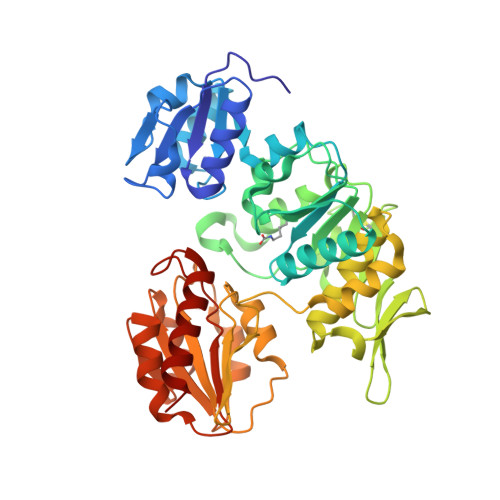
    Mur ligases have essential roles in the biosynthesis of peptidoglycan, and they represent attractive targets for the design of novel antibacterials. MurD (UDP-N-acetylmuramoyl-L-alanine:D-glutamate ligase) is the second enzyme in the series of Mur ligases, and it catalyzes the addition of D-glutamic acid (D-Glu) to the cytoplasmic intermediate UDP-N-acetylmuramoyl-L-alanine (UMA). Because of the high binding affinity of D-Glu toward MurD, we synthesized and biochemically evaluated a series of N-substituted D-Glu derivatives as potential inhibitors of MurD from E. coli, which allowed us to explore the structure-activity relationships.The substituted naphthalene-N-sulfonyl-D-Glu inhibitors, which were synthesized as potential transition state analogues, displayed IC50 values ranging from 80 to 600 microM. In addition, the high-resolution crystal structures of MurD in complex with four novel inhibitors revealed details of the binding mode of the inhibitors within the active site of MurD. Structure-activity relationships and cocrystal structures constitute an excellent starting point for further development of novel MurD inhibitors of this structural class.


  • Organizational Affiliation
    • Drug Discovery, Lek Pharmaceuticals d.d., Verovskova 57, 1526 Ljubljana, Slovenia.

Macromolecule Content 

  • Total Structure Weight: 48.74 kDa 
  • Atom Count: 3,777 
  • Modeled Residue Count: 440 
  • Deposited Residue Count: 445 
  • Unique protein chains: 1

Macromolecules

Find similar proteins by:|  3D Structure
Entity ID: 1
MoleculeChains  Sequence LengthOrganismDetailsImage
UDP-N-ACETYLMURAMOYLALANINE--D-GLUTAMATE LIGASE445Escherichia coliMutation(s): 0 
EC: 6.3.2.9
UniProt
Find proteins for P14900 (Escherichia coli (strain K12))
Explore P14900 
Go to UniProtKB:  P14900
Entity Groups
Sequence Clusters30% Identity50% Identity70% Identity90% Identity95% Identity100% Identity
UniProt GroupP14900
Sequence Annotations
Expand
Reference Sequence

Experimental Data & Validation

Experimental Data

  • Method: X-RAY DIFFRACTION
  • Resolution: 1.88 Å
  • R-Value Free:  0.222 (Depositor), 0.224 (DCC) 
  • R-Value Work:  0.183 (Depositor), 0.183 (DCC) 
  • R-Value Observed: 0.184 (Depositor) 
Space Group: P 41
Unit Cell:
Length ( Å )Angle ( ˚ )
a = 65.517α = 90
b = 65.517β = 90
c = 136.174γ = 90
Software Package:
Software NamePurpose
REFMACrefinement
XDSdata reduction
XSCALEdata scaling
PHASERphasing

Structure Validation

View Full Validation Report


Ligand Structure Quality Assessment 


Entry History 

Deposition Data

Revision History  (Full details and data files)

  • Version 1.0: 2008-03-25
    Type: Initial release
  • Version 1.1: 2011-05-08
    Changes: Version format compliance
  • Version 1.2: 2011-07-13
    Changes: Version format compliance
  • Version 1.3: 2018-01-31
    Changes: Database references, Source and taxonomy
  • Version 1.4: 2023-12-13
    Changes: Data collection, Database references, Derived calculations, Other, Refinement description