X-ray crystallographic studies on hexameric insulins in the presence of helix-stabilizing agents, thiocyanate, methylparaben, and phenol.
Whittingham, J.L., Chaudhuri, S., Dodson, E.J., Moody, P.C., Dodson, G.G.(1995) Biochemistry 34: 15553-15563
- PubMed: 7492558
- DOI: https://doi.org/10.1021/bi00047a022
- Primary Citation of Related Structures:
1MPJ, 2TCI, 3MTH - PubMed Abstract:
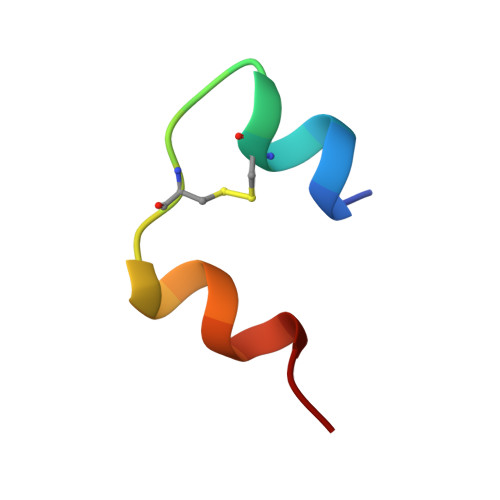
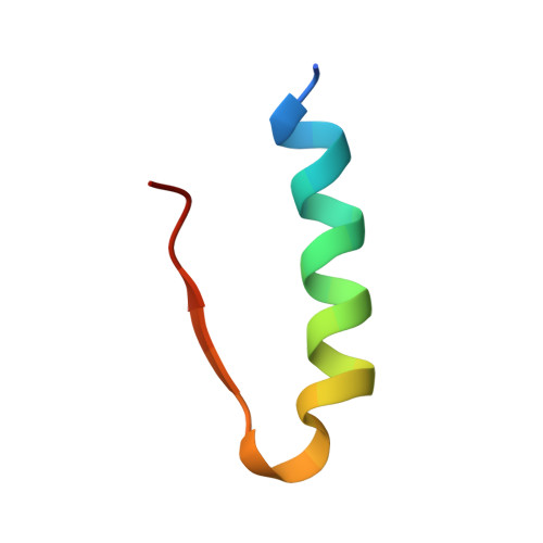
Three X-ray crystallographic studies have been carried out on pig insulin in the presence of three ligands, thiocyanate, methylparaben (methyl p-hydroxybenzoate), and phenol. In each case, rhombohedral crystals were obtained, which diffracted to 1.8, 1.9, and 2.3 A, respectively. Each crystal structure was very similar to that of 4-zinc pig insulin, which was used as a starting model for PROLSQ refinement (Collaborative Computational Project, Number 4, 1994). The R factors for the refined structures of thiocyanate insulin, methylparaben insulin, and phenol insulin were 19.6, 18.4, and 19.1, respectively. Each crystal structure consists of T3R3f insulin hexamers with two zinc ions per hexamer. In the R3f trimer of the thiocyanate insulin hexamer, one thiocyanate ion is coordinated to the zinc on the hexamer 3-fold axis, but there is no evidence of zinc ion binding in the off-axis zinc ion sites seen in the 4-zinc pig insulin structure. In the methylparaben insulin and phenol insulin hexamers, the phenolic ligands are bound at the dimer-dimer interfaces in the R3f trimers in a manner similar to that of phenol in R6 phenol insulin. The binding of methylparaben appears to make the hexamer more compact by drawing the A and the B chains closer together in the binding site. In all three structures presented herein, the conformations of the first three residues of the B chain in the R3f trimer are extended rather than alpha-helical, as is seen in R6 phenol insulin. The energetics of ligand binding in the insulin hexamer are discussed.
Organizational Affiliation:
Department of Chemistry, University of York, Heslington, England.


