


The solution structure of membrane protein
Kuwahara, Y., Unzai, S., Nagata, T., Hiroaki, H.To be published.
Experimental Data Snapshot
wwPDB Validation 3D Report Full Report

Entity ID: 1 | |||||
|---|---|---|---|---|---|
| Molecule | Chains | Sequence Length | Organism | Details | Image |
| hypothetical membrane protein | 113 | Pyrococcus horikoshii | Mutation(s): 0 Gene Names: PH0470 | 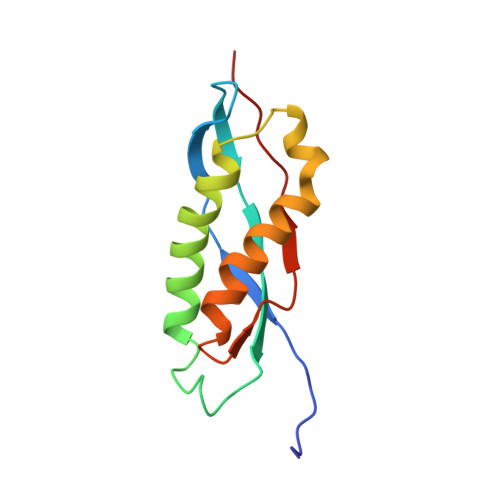 | |
UniProt | |||||
Entity Groups | |||||
| Sequence Clusters | 30% Identity50% Identity70% Identity90% Identity95% Identity100% Identity | ||||
| UniProt Group | O58205 | ||||
Sequence AnnotationsExpand | |||||
Reference Sequence | |||||


