2RCZ | pdb_00002rcz

Structure of the second PDZ domain of ZO-1


Experimental Data Snapshot

  • Method: X-RAY DIFFRACTION
  • Resolution: 1.70 Å
  • R-Value Free: 
    0.258 (Depositor), 0.260 (DCC) 
  • R-Value Work: 
    0.204 (Depositor), 0.204 (DCC) 
  • R-Value Observed: 
    0.209 (Depositor) 

wwPDB Validation 3D Report Full Report

Validation slider image for 2RCZ

This is version 1.2 of the entry. See complete history

Literature

Domain swapping within PDZ2 is responsible for dimerization of ZO proteins.

Fanning, A.S.Lye, M.F.Anderson, J.M.Lavie, A.

(2007) J Biological Chem 282: 37710-37716

  • DOI: https://doi.org/10.1074/jbc.M707255200
  • Primary Citation Related Structures: 
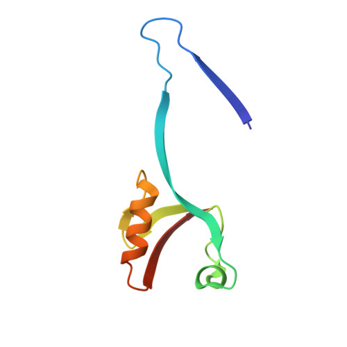
    2RCZ

  • PubMed Abstract: 

    ZO-1 is a multidomain protein involved in cell-cell junctions and contains three PDZ domains, which are necessary for its function in vivo. PDZ domains play a central role in assembling diverse protein complexes through their ability to recognize short peptide motifs on other proteins. We determined the structure of the second of the three PDZ domains of ZO-1, which is known to promote dimerization as well as bind to C-terminal sequences on connexins. The dimer is stabilized by extensive symmetrical domain swapping of beta-strands, which is unlike any other known mechanism of PDZ dimerization. The canonical peptide-binding groove remains intact in both subunits of the PDZ2 dimer and is created by elements contributed from both monomers. This unique structure reveals an additional example of how PDZ domains dimerize and has multiple implications for both peptide binding and oligomerization in vivo.


  • Organizational Affiliation
    • Department of Cell and Molecular Physiology, University of North Carolina at Chapel Hill, Chapel Hill, NC 27599-7545, USA.

Macromolecule Content 

  • Total Structure Weight: 18.01 kDa 
  • Atom Count: 1,302 
  • Modeled Residue Count: 160 
  • Deposited Residue Count: 162 
  • Unique protein chains: 1

Macromolecules

Find similar proteins by:|  3D Structure
Entity ID: 1
MoleculeChains  Sequence LengthOrganismDetailsImage
Tight junction protein ZO-1
A, B
81Homo sapiensMutation(s): 0 
Gene Names: TJP1ZO1
UniProt & NIH Common Fund Data Resources
Find proteins for Q07157 (Homo sapiens)
Explore Q07157 
Go to UniProtKB:  Q07157
PHAROS:  Q07157
GTEx:  ENSG00000104067 
Entity Groups
Sequence Clusters30% Identity50% Identity70% Identity90% Identity95% Identity100% Identity
UniProt GroupQ07157
Sequence Annotations
Expand
Reference Sequence

Experimental Data & Validation

Experimental Data

  • Method: X-RAY DIFFRACTION
  • Resolution: 1.70 Å
  • R-Value Free:  0.258 (Depositor), 0.260 (DCC) 
  • R-Value Work:  0.204 (Depositor), 0.204 (DCC) 
  • R-Value Observed: 0.209 (Depositor) 
Space Group: C 1 2 1
Unit Cell:
Length ( Å )Angle ( ˚ )
a = 47.75α = 90
b = 33.61β = 103.64
c = 91.04γ = 90
Software Package:
Software NamePurpose
XSCALEdata scaling
PHASERphasing
REFMACrefinement
PDB_EXTRACTdata extraction

Structure Validation

View Full Validation Report


Entry History 

Deposition Data

Revision History  (Full details and data files)

  • Version 1.0: 2007-10-09
    Type: Initial release
  • Version 1.1: 2011-07-13
    Changes: Advisory, Version format compliance
  • Version 1.2: 2024-02-21
    Changes: Data collection, Database references