2RA6 | pdb_00002ra6

Crystal Structure of the Possum Milk Whey Lipocalin Trichosurin at pH 4.6 with Bound 4-ethylphenol


Experimental Data Snapshot

  • Method: X-RAY DIFFRACTION
  • Resolution: 1.50 Å
  • R-Value Free: 
    0.241 (Depositor), 0.233 (DCC) 
  • R-Value Work: 
    0.226 (Depositor), 0.226 (DCC) 

Starting Model: experimental
View more details

wwPDB Validation 3D Report Full Report

Validation slider image for 2RA6

This is version 1.4 of the entry. See complete history

Literature

Three-dimensional structure and ligand binding properties of trichosurin, a metatherian lipocalin from the milk whey of the common brushtail possum Trichosurus vulpecula

Watson, R.P.Demmer, J.Baker, E.N.Arcus, V.L.

(2007) Biochem J 408: 29-38

  • DOI: https://doi.org/10.1042/BJ20070567
  • Primary Citation Related Structures: 
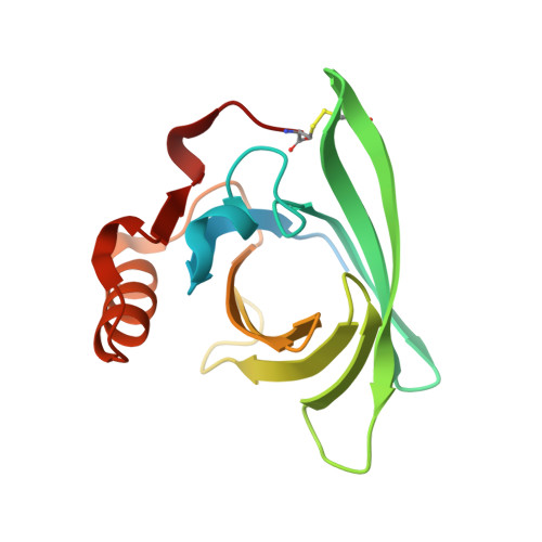
    2R73, 2R74, 2RA6

  • PubMed Abstract: 

    Lipocalins are extracellular proteins (17-25 kDa) that bind and transport small lipophilic molecules. The three-dimensional structure of the first lipocalin from a metatherian has been determined at different values of pH both with and without bound ligands. Trichosurin, a protein from the milk whey of the common brushtail possum, Trichosurus vulpecula, has been recombinantly expressed in Escherichia coli, refolded from inclusion bodies, purified and crystallized at two different pH values. The three-dimensional structure of trichosurin was solved by X-ray crystallography in two different crystal forms to 1.9 A (1 A=0.1 nm) and 2.6 A resolution, from crystals grown at low and high pH values respectively. Trichosurin has the typical lipocalin fold, an eight-stranded anti-parallel beta-barrel but dimerizes in an orientation that has not been seen previously. The putative binding pocket in the centre of the beta-barrel is well-defined in both high and low pH structures and is occupied by water molecules along with isopropanol molecules from the crystallization medium. Trichosurin was also co-crystallized with a number of small molecule ligands and structures were determined with 2-naphthol and 4-ethylphenol bound in the centre of the beta-barrel. The binding of phenolic compounds by trichosurin provides clues to the function of this important marsupial milk protein, which is highly conserved across metatherians.


  • Organizational Affiliation
    • Laboratory of Structural Biology, School of Biological Sciences, University of Auckland, Private Bag 92-019, Auckland, New Zealand.

Macromolecule Content 

  • Total Structure Weight: 80.52 kDa 
  • Atom Count: 5,187 
  • Modeled Residue Count: 569 
  • Deposited Residue Count: 664 
  • Unique protein chains: 1

Macromolecules

Find similar proteins by:|  3D Structure
Entity ID: 1
MoleculeChains  Sequence LengthOrganismDetailsImage
Trichosurin
A, B, C, D
166Trichosurus vulpeculaMutation(s): 1 
UniProt
Find proteins for Q29147 (Trichosurus vulpecula)
Explore Q29147 
Go to UniProtKB:  Q29147
Entity Groups
Sequence Clusters30% Identity50% Identity70% Identity90% Identity95% Identity100% Identity
UniProt GroupQ29147
Sequence Annotations
Expand
Reference Sequence

Small Molecules

Ligands 4 Unique
IDChains Name / Formula / InChI Key2D Diagram3D Interactions
ETY

Query on ETY


Download:Ideal Coordinates CCD File
CA [auth D],
H [auth A],
P [auth B],
V [auth C]
4-ethylphenol
C8 H10 O
HXDOZKJGKXYMEW-UHFFFAOYSA-N
ZN

Query on ZN


Download:Ideal Coordinates CCD File
E [auth A]
K [auth B]
L [auth B]
S [auth C]
Y [auth D]
E [auth A],
K [auth B],
L [auth B],
S [auth C],
Y [auth D],
Z [auth D]
ZINC ION
Zn
PTFCDOFLOPIGGS-UHFFFAOYSA-N
IPA

Query on IPA


Download:Ideal Coordinates CCD File
DA [auth D]
EA [auth D]
I [auth A]
J [auth A]
Q [auth B]
DA [auth D],
EA [auth D],
I [auth A],
J [auth A],
Q [auth B],
R [auth B],
W [auth C],
X [auth C]
ISOPROPYL ALCOHOL
C3 H8 O
KFZMGEQAYNKOFK-UHFFFAOYSA-N
CL

Query on CL


Download:Ideal Coordinates CCD File
AA [auth D]
BA [auth D]
F [auth A]
G [auth A]
M [auth B]
AA [auth D],
BA [auth D],
F [auth A],
G [auth A],
M [auth B],
N [auth B],
O [auth B],
T [auth C],
U [auth C]
CHLORIDE ION
Cl
VEXZGXHMUGYJMC-UHFFFAOYSA-M

Experimental Data & Validation

Experimental Data

  • Method: X-RAY DIFFRACTION
  • Resolution: 1.50 Å
  • R-Value Free:  0.241 (Depositor), 0.233 (DCC) 
  • R-Value Work:  0.226 (Depositor), 0.226 (DCC) 
Space Group: P 1 21 1
Unit Cell:
Length ( Å )Angle ( ˚ )
a = 44.318α = 90
b = 100.47β = 90.02
c = 84.702γ = 90
Software Package:
Software NamePurpose
AMoREphasing
CNSrefinement
XSCALEdata scaling

Structure Validation

View Full Validation Report


Entry History 

Deposition Data

  • Released Date: 2007-11-20 
  • Deposition Author(s): Watson, R.P.

Revision History  (Full details and data files)

  • Version 1.0: 2007-11-20
    Type: Initial release
  • Version 1.1: 2011-07-13
    Changes: Version format compliance
  • Version 1.2: 2021-11-10
    Changes: Database references, Derived calculations
  • Version 1.3: 2023-10-25
    Changes: Data collection, Refinement description
  • Version 1.4: 2024-10-16
    Changes: Structure summary