2RA0 | pdb_00002ra0

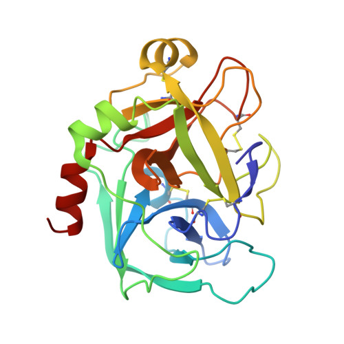
X-ray Structure of FXa in complex with 7-fluoroindazole


Experimental Data Snapshot

  • Method: X-RAY DIFFRACTION
  • Resolution: 2.30 Å
  • R-Value Free: 
    0.314 (Depositor), 0.303 (DCC) 
  • R-Value Work: 
    0.246 (Depositor), 0.238 (DCC) 

wwPDB Validation 3D Report Full Report

Validation slider image for 2RA0

Ligand Structure Quality Assessment 


This is version 1.3 of the entry. See complete history. 

Literature

7-fluoroindazoles as potent and selective inhibitors of factor xa.

Lee, Y.K., Parks, D.J., Lu, T., Thieu, T.V., Markotan, T., Pan, W., McComsey, D.F., Milkiewicz, K.L., Crysler, C.S., Ninan, N., Abad, M.C., Giardino, E.C., Maryanoff, B.E., Damiano, B.P., Player, M.R.

(2008) J Med Chem 51: 282-297

  • DOI: https://doi.org/10.1021/jm701217r
  • Primary Citation Related Structures: 
    2RA0

  • PubMed Abstract: 

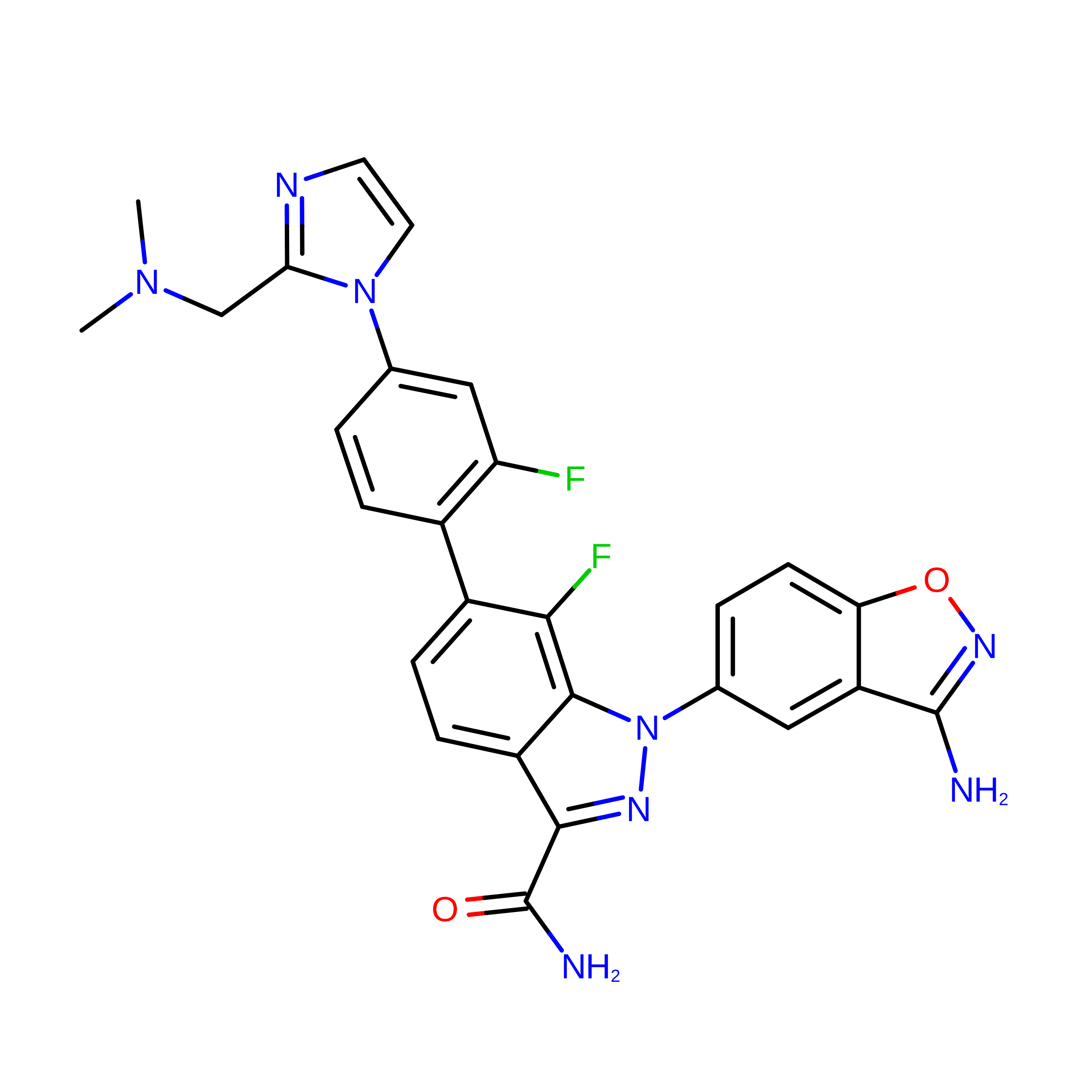
    We have developed a novel series of potent and selective factor Xa inhibitors that employ a key 7-fluoroindazolyl moiety. The 7-fluoro group on the indazole scaffold replaces the carbonyl group of an amide that is found in previously reported factor Xa inhibitors. The structure of a factor Xa cocrystal containing 7-fluoroindazole 51a showed the 7-fluoro atom hydrogen-bonding with the N-H of Gly216 (2.9 A) in the peptide backbone. Thus, the 7-fluoroindazolyl moiety not only occupied the same space as the carbonyl group of an amide found in prior factor Xa inhibitors but also maintained a hydrogen bond interaction with the protein's beta-sheet domain. The structure-activity relationship for this series was consistent with this finding, as the factor Xa inhibitory potencies were about 60-fold greater (DeltaDelta G approximately 2.4 kcal/mol) for the 7-fluoroindazoles 25a and 25c versus the corresponding indazoles 25b and 25d. Highly convergent synthesis of these factor Xa inhibitors is also described.


  • Organizational Affiliation: 
    • Johnson & Johnson Pharmaceutical Research and Development, Welsh and McKean Roads, Spring House, Pennsylvania 19477-0776, USA.

Macromolecule Content 

  • Total Structure Weight: 32.42 kDa 
  • Atom Count: 2,348 
  • Modeled Residue Count: 285 
  • Deposited Residue Count: 285 
  • Unique protein chains: 2

Macromolecules

Find similar proteins by:|  3D Structure
Entity ID: 1
MoleculeChains  Sequence LengthOrganismDetailsImage
Coagulation factor X234Homo sapiensMutation(s): 0 
Gene Names: F10
EC: 3.4.21.6
UniProt & NIH Common Fund Data Resources
Find proteins for P00742 (Homo sapiens)
Explore P00742 
Go to UniProtKB:  P00742
PHAROS:  P00742
GTEx:  ENSG00000126218 
Entity Groups
Sequence Clusters30% Identity50% Identity70% Identity90% Identity95% Identity100% Identity
UniProt GroupP00742
Sequence Annotations
Expand
Reference Sequence
Find similar proteins by:|  3D Structure
Entity ID: 2
MoleculeChains  Sequence LengthOrganismDetailsImage
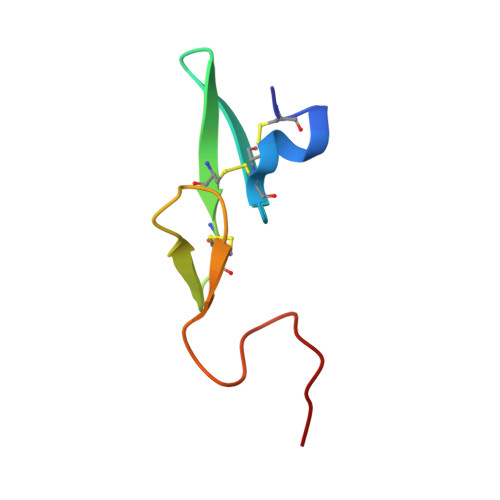
Coagulation factor XB [auth L]51Homo sapiensMutation(s): 1 
Gene Names: F10
EC: 3.4.21.6
UniProt & NIH Common Fund Data Resources
Find proteins for P00742 (Homo sapiens)
Explore P00742 
Go to UniProtKB:  P00742
PHAROS:  P00742
GTEx:  ENSG00000126218 
Entity Groups
Sequence Clusters30% Identity50% Identity70% Identity90% Identity95% Identity100% Identity
UniProt GroupP00742
Sequence Annotations
Expand
Reference Sequence

Small Molecules

Ligands 1 Unique
IDChains Name / Formula / InChI Key2D Diagram3D Interactions
JNJ

Query on JNJ


Download:Ideal Coordinates CCD File
C [auth A]1-(3-amino-1,2-benzisoxazol-5-yl)-6-(4-{2-[(dimethylamino)methyl]-1H-imidazol-1-yl}-2-fluorophenyl)-7-fluoro-1H-indazole-3-carboxamide
C27 H22 F2 N8 O2
MGUDDBRJHXFTEY-UHFFFAOYSA-N

Experimental Data & Validation

Experimental Data

  • Method: X-RAY DIFFRACTION
  • Resolution: 2.30 Å
  • R-Value Free:  0.314 (Depositor), 0.303 (DCC) 
  • R-Value Work:  0.246 (Depositor), 0.238 (DCC) 
Space Group: P 21 21 21
Unit Cell:
Length ( Å )Angle ( ˚ )
a = 48.871α = 90
b = 74.984β = 90
c = 75.089γ = 90
Software Package:
Software NamePurpose
SAINTdata scaling
CNSrefinement
PDB_EXTRACTdata extraction
PROTEUM PLUSdata collection
SAINTdata reduction
CNSphasing

Structure Validation

View Full Validation Report


Ligand Structure Quality Assessment 


Entry History 

Deposition Data

  • Released Date: 2008-01-29 
  • Deposition Author(s): Abad, M.C.

Revision History  (Full details and data files)

  • Version 1.0: 2008-01-29
    Type: Initial release
  • Version 1.1: 2011-07-13
    Changes: Version format compliance
  • Version 1.2: 2021-10-20
    Changes: Database references, Derived calculations
  • Version 1.3: 2024-10-30
    Changes: Data collection, Structure summary