2R6S | pdb_00002r6s

Crystal structure of Gab protein


Experimental Data Snapshot

  • Method: X-RAY DIFFRACTION
  • Resolution: 2.10 Å
  • R-Value Free: 
    0.199 (Depositor), 0.208 (DCC) 
  • R-Value Work: 
    0.162 (Depositor), 0.170 (DCC) 
  • R-Value Observed: 
    0.163 (Depositor) 

Starting Model: experimental
View more details

wwPDB Validation 3D Report Full Report

Validation slider image for 2R6S

Ligand Structure Quality Assessment 


This is version 1.3 of the entry. See complete history

Literature

A mixture of fortunes: the curious determination of the structure of Escherichia coli BL21 Gab protein.

Lohkamp, B.Dobritzsch, D.

(2008) Acta Crystallogr D Biol Crystallogr 64: 407-415

  • DOI: https://doi.org/10.1107/S0907444908001091
  • Primary Citation Related Structures: 
    2R6S

  • PubMed Abstract: 

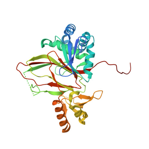
    In protein crystallography, monodisperse protein samples of high purity are usually required in order to obtain diffraction-quality crystals. Here, crystals were reproducibly grown from a protein sample before its homogeneity had been determined. The sample was obtained after the first attempt to purify a recombinant target protein from an Escherichia coli cell lysate. Subsequent analysis revealed that it was a mixture of about 50 different proteins with no predominant species. Diffraction data were collected to 2.1 A and the space group was identified as I422. A molecular-replacement search with models of the expected target did not give a solution, which suggested that a contaminating E. coli protein had been crystallized. A PDB search revealed 256 structures determined in space group I422, of which 14 are E. coli proteins and two have unit-cell parameters similar to those observed. Molecular replacement with these structures showed a clear solution for one of them, the Gab protein. The structure is presented and compared with the deposited structure, from which it shows small but significant differences. The refined model contains bicine and sulfate as bound ligands, which provide insights into possible substrate-binding sites.


  • Organizational Affiliation
    • Department of Medical Biochemistry and Biophysics, Karolinska Institutet, Sweden.

Macromolecule Content 

  • Total Structure Weight: 39.86 kDa 
  • Atom Count: 2,938 
  • Modeled Residue Count: 299 
  • Deposited Residue Count: 325 
  • Unique protein chains: 1

Macromolecules

Find similar proteins by:|  3D Structure
Entity ID: 1
MoleculeChains  Sequence LengthOrganismDetailsImage
Gab protein325Escherichia coliMutation(s): 1 

Small Molecules

Ligands 4 Unique
IDChains Name / Formula / InChI Key2D Diagram3D Interactions
BCN

Query on BCN


Download:Ideal Coordinates CCD File
F [auth A]BICINE
C6 H13 N O4
FSVCELGFZIQNCK-UHFFFAOYSA-N
SO4

Query on SO4


Download:Ideal Coordinates CCD File
C [auth A],
D [auth A],
E [auth A]
SULFATE ION
O4 S
QAOWNCQODCNURD-UHFFFAOYSA-L
GOL

Query on GOL


Download:Ideal Coordinates CCD File
AA [auth A]
G [auth A]
H [auth A]
I [auth A]
J [auth A]
AA [auth A],
G [auth A],
H [auth A],
I [auth A],
J [auth A],
K [auth A],
L [auth A],
M [auth A],
N [auth A],
O [auth A],
P [auth A],
Q [auth A],
R [auth A],
S [auth A],
T [auth A],
U [auth A],
V [auth A],
W [auth A],
X [auth A],
Y [auth A],
Z [auth A]
GLYCEROL
C3 H8 O3
PEDCQBHIVMGVHV-UHFFFAOYSA-N
FE2

Query on FE2


Download:Ideal Coordinates CCD File
B [auth A]FE (II) ION
Fe
CWYNVVGOOAEACU-UHFFFAOYSA-N

Experimental Data & Validation

Experimental Data

  • Method: X-RAY DIFFRACTION
  • Resolution: 2.10 Å
  • R-Value Free:  0.199 (Depositor), 0.208 (DCC) 
  • R-Value Work:  0.162 (Depositor), 0.170 (DCC) 
  • R-Value Observed: 0.163 (Depositor) 
Space Group: I 4 2 2
Unit Cell:
Length ( Å )Angle ( ˚ )
a = 120.9α = 90
b = 120.9β = 90
c = 137.17γ = 90
Software Package:
Software NamePurpose
REFMACrefinement
PDB_EXTRACTdata extraction
ProDCdata collection
MOSFLMdata reduction
SCALAdata scaling
MOLREPphasing

Structure Validation

View Full Validation Report


Ligand Structure Quality Assessment 


Entry History 

Deposition Data

Revision History  (Full details and data files)

  • Version 1.0: 2008-06-03
    Type: Initial release
  • Version 1.1: 2011-07-13
    Changes: Non-polymer description, Version format compliance
  • Version 1.2: 2023-02-08
    Changes: Database references, Derived calculations, Source and taxonomy
  • Version 1.3: 2023-09-20
    Changes: Data collection, Refinement description