2R65 | pdb_00002r65

Crystal structure of Helicobacter pylori ATP dependent protease, FtsH ADP complex


Experimental Data Snapshot

  • Method: X-RAY DIFFRACTION
  • Resolution: 3.30 Å
  • R-Value Free: 
    0.331 (Depositor), 0.312 (DCC) 
  • R-Value Work: 
    0.288 (Depositor), 0.269 (DCC) 
  • R-Value Observed: 
    0.290 (Depositor) 

Starting Model: experimental
View more details

wwPDB Validation 3D Report Full Report

Validation slider image for 2R65

Ligand Structure Quality Assessment 


This is version 1.3 of the entry. See complete history

Literature

Structural studies on Helicobacter pyloriATP-dependent protease, FtsH

Kim, S.H.Kang, G.B.Song, H.-E.Park, S.J.Bea, M.-H.Eom, S.H.

(2008) J Synchrotron Radiat 15: 208-210

  • DOI: https://doi.org/10.1107/S090904950706846X
  • Primary Citation Related Structures: 
    2R62, 2R65

  • PubMed Abstract: 

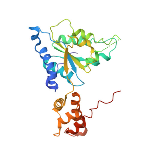
    The ATP-dependent protease, FtsH, degrades misassembled membrane proteins for quality control like SecY, subunit a of FoF1-ATPase, and YccA, and digests short-lived soluble proteins in order to control their cellular regulation, including sigma32, LpxC and lambdacII. The FtsH protein has an N-terminal transmembrane segment and a large cytosolic region that consists of two domains, an ATPase and a protease domain. To provide a structural basis for the nucleotide-dependent domain motions and a better understanding of substrate translocation, the crystal structures of the Helicobacter pylori (Hp) FtsH ATPase domain in the nucleotide-free state and complexed with ADP, were determined. Two different structures of HpFtsH ATPase were observed, with the nucleotide-free state in an asymmetric unit, and these structures reveal the new forms and show other conformational differences between the nucleotide-free and ADP-bound state compared with previous structures. In particular, one HpFtsH Apo structure has a considerable rotation difference compared with the HpFtsH ADP complex, and this large conformational change reveals that FtsH may have the mechanical force needed for substrate translocation.


  • Organizational Affiliation
    • Department of Life Science, Cell Dynamics Research Center, Gwangju Institute of Science and Technology, Gwangju 500-712, Korea.

Macromolecule Content 

  • Total Structure Weight: 147.68 kDa 
  • Atom Count: 9,645 
  • Modeled Residue Count: 1,245 
  • Deposited Residue Count: 1,340 
  • Unique protein chains: 1

Macromolecules

Find similar proteins by:|  3D Structure
Entity ID: 1
MoleculeChains  Sequence LengthOrganismDetailsImage
Cell division protease ftsH homolog
A, B, C, D, E
268Helicobacter pyloriMutation(s): 1 
Gene Names: ftsH
EC: 3.4.24
UniProt
Find proteins for P71408 (Helicobacter pylori (strain ATCC 700392 / 26695))
Explore P71408 
Go to UniProtKB:  P71408
Entity Groups
Sequence Clusters30% Identity50% Identity70% Identity90% Identity95% Identity100% Identity
UniProt GroupP71408
Sequence Annotations
Expand
Reference Sequence

Experimental Data & Validation

Experimental Data

  • Method: X-RAY DIFFRACTION
  • Resolution: 3.30 Å
  • R-Value Free:  0.331 (Depositor), 0.312 (DCC) 
  • R-Value Work:  0.288 (Depositor), 0.269 (DCC) 
  • R-Value Observed: 0.290 (Depositor) 
Space Group: C 2 2 21
Unit Cell:
Length ( Å )Angle ( ˚ )
a = 110.471α = 90
b = 219.485β = 90
c = 146.692γ = 90
Software Package:
Software NamePurpose
REFMACrefinement
HKL-2000data collection
HKL-2000data reduction
HKL-2000data scaling
PHASESphasing

Structure Validation

View Full Validation Report


Ligand Structure Quality Assessment 


Entry History 

Deposition Data

Revision History  (Full details and data files)

  • Version 1.0: 2008-09-09
    Type: Initial release
  • Version 1.1: 2011-07-13
    Changes: Advisory, Version format compliance
  • Version 1.2: 2021-11-10
    Changes: Database references, Derived calculations
  • Version 1.3: 2023-10-25
    Changes: Data collection, Refinement description